Quantcast
Channel: Clinical – ALiEM
Viewing all 273 articles
Browse latest View live

Did the Affordable Care Act actually reduce ED visits as politicians promised?

$
0
0

ACA

The Affordable Care Act (ACA) was supposed to expand coverage to the uninsured and many politicians claimed this would result in lower use of “expensive emergency rooms” for the treatment of patients’ acute complaints. Ignore, for the moment, the controversy about whether or not the emergency department (ED) is an expensive or appropriate place for patients to seek care. A new survey [PDF] from the Center for Disease Control and Prevention (CDC), asked the question: Did the ACA actually reduce ED visits as politicians promised [1]?

Background

This presumption, that increased health insurance coverage would decrease ED visits, runs counter to 2 facts.

  1. Only one-fifth of patients visit the ED due to problems such as their doctor’s office being closed or lack of other providers. The vast majority goes to the ED because they (or any other prudent layperson) think they are having an actual emergency.
  2. recent randomized experiment demonstrated that giving Medicaid to the uninsured in Oregon led to a 40% increase in ED visits [2].

The CDC study did not answer the question about ED visit reduction

The authors used the National Health Interview Survey to assess patterns of ED use among adults age 18-64 in 2013 and 2014 — the years straddling the ACA’s major insurance expansions [1].

Figure 1 ACA - no label

Fig 1. Adults (age 18-64) with ≥1 visit to the ED in the past year, by number of visits, health insurance coverage status, and year: United States (2013 and 2014). Figure from [1].

In this study based on patient recollection, the percentage of people reporting an ED visit during the previous 12 months did not significantly differ between 2013 and 2014. This is true for both the Medicaid and privately insured subgroups. We know that about 10 million people gained insurance coverage due to the ACA’s expansion of Medicaid and through private health insurance marketplaces in 2014. How is it that none of those people ventured their way into an ED?

Contrast these findings to the highly cited Oregon Medicaid experiment clearly demonstrated that the absolute number of total ED visits went up by about 40% during its Medicaid expansion at 18 months [2]. This Oregon study also showed that the percent of patients with any visit, which is the same measure reported by CDC, also significantly increased by about 20%.

So why are the CDC and Oregon studies not in agreement?

Like any other scientific paper, the devil is in the details, or in academic-speak, the methods. The Oregon study results differ from the CDC study in that the former used an administrative database and the latter used patient self-report. A true comparison would require comparing the CDC data to self-reported ED visits. Fortunately, the Oregon researchers provided us that information as well (Figure 2). The in-person interview at 12 months showed no significant difference for patients getting Medicaid on ED visits.


Figure 2 ACA copy

Fig 2. Comparing ED visit results from administrative data and self-reports. Table from [2]

So ultimately in the end, both the Oregon and the CDC study – looking at a 12 month period – found that patients did not report any significant change in the likelihood of visiting an ED [1,2]. So where did this 40% increase in ED visits in Oregon come from? If you look at Figure 2, the administrative ED data at 18 months clearly show significant increases in percentage of patients with any ED visit and the total number of ED visits.

So maybe it’s true that Obamacare nationally hasn’t changed the overall number or likelihood of visiting the ED – which runs contrary to our predictions – but that might just be the results of the sampling method (self-report vs. administrative database) or too short an observation period (12 vs 18 months).

I would be willing to bet that when researchers look at the real numbers of how many patients actually went to the ED, not just what interviewees say they did (which is subject to recall bias), our predictions of a spike in ED visits will be corroborated.

Bottom line

To honestly answer the question – Did the ACA actually reduce ED visits as politicians promised? This CDC study did not clearly answer the question. What we need is a true analysis of the administrative records, free from recall bias.


References

  1. Gindi RM, Black LI, Cohen RA. Reasons for Emergency Room Use Among U.S. Adults Aged 18-64: National Health Interview Survey, 2013 and 2014. Natl Health Stat Report. 2016; (90):1-16. PMID: 26905514
  2. Taubman SL, Allen HL, Wright BJ, Baicker K, Finkelstein AN. Medicaid increases emergency-department use: evidence from Oregon’s Health Insurance Experiment. Science. 2014; 343(6168):263-8. PMID: 24385603

Author information

Cedric Dark, MD MPH

Cedric Dark, MD MPH

Assistant Professor of Medicine
Section of Emergency Medicine
Baylor College of Medicine;
Founder & Executive Editor,
Policy Prescriptions ® (www.policyrx.org)

The post Did the Affordable Care Act actually reduce ED visits as politicians promised? appeared first on ALiEM.


Synthetic cannabinoid use reaches new heights: K2 is not just a mountain on the China-Pakistan boarder

$
0
0

K2You are working a shift in the emergency department, and you hear the ambulance sirens. EMS is bringing you two patients, friends from a nearby shelter. Per report, the two men were “smoking drugs” together outside of the shelter. Bystanders noted that the 29-year-old man became increasingly agitated, shouting, banging on the door, and threatening his other shelter mates, while the other, a 50-year-old man, laid down on the sidewalk. EMS also reports picking up these patients in an area known for high “K2” use.

Synthetic Cannabinoids: Background

Synthetic cannabinoids (SC), sometimes called synthetic cannabinoid receptor agonists (SCRA), were originally developed to allow investigation of the human endocannabinoid receptor system. As more was learned about this endogenous system, interest grew in their use as therapeutic drugs for various medical ailments, such as the alleviation of the gastrointestinal side effects of chemotherapeutic agents. These compounds interact with the cannabinoid (CB) receptors as does delta-9-tetrahydrocannabinol (THC), the psychoactive component in Cannabis, but they are structurally different and generally more potent [1-3].

Currently, SCs can be classified into several different types: [1, 4]

  • Aminoalkylindoles
  • Cyclohexylphenols
  • Benzylindoles
  • THC analogues (also referred to as classic synthetic cannabinoids)

Synthetic chemists, primarily in China, can create hundreds of different SCs, and this helps account for the unpredictable clinical effects seen in the ED. The HBO video below elaborates about the production of SCs in China.

Growing popularity of SC’s

SCs entered the “black market” in 2004 as a novel class of designer drugs marketed with names such as “Spice” and “K2.” Since then, they have grown in popularity throughout Europe and North America. Initially SCs were sold legally (“hidden in plain sight”) as an alternative to Cannabis and they became readily available in gas stations, head shops, and over the Internet. Because of the chemicals’ high potencies and small volumes needed, they are often sprayed onto a bulky substance (e.g., crushed dried plant matter) that resembles Cannabis to allow for easy delivery. It is impossible to predict the dose of SCs associated with any product mainly because of the method in which they are produced (i.e. the bulky plant matter can have any amount of SCs or none at all). The average cost in 2010 was approximately $20-30 per 3 grams of herbal material [5]. In 2012, the Monitoring the Future survey on adolescent drug use reported that about 12% of 12th-graders use SCs [6].

Despite legal efforts since 2011 to ban these substances, SC use has skyrocketed in urban areas, predominantly among the homeless population. Its falling price, as low as five dollars per “bag” containing from four to ten grams, its association with a “stronger high” when compared to Cannabis, and its easy accessibility all add to its popularity [3, 7]. With recent seizures in small shops in New York City yielding over two million packages, the supply of SCs appears to be so vast that this may account for its dropping price. In addition, SCs’ inconspicuous nature (i.e they are not detected by standard drug testing for THC) made them popular in schools and among athletes.

This New York Times article depicts the more current use of SCs in east Harlem [7].

Can SC’s be easily detected in the urine today?

The answer is still NO. The most common urine drug screen still used today is the EMIT, or enzyme mediated immunoassay technique, specifically designed to detect a chemical structure present above a specified cutoff of detection. Currently, some assays screen for “cannabinoid” but typically only detect THC-COOH (a metabolite of THC) at concentrations greater only than 50 ng/mL. At best, newer urine drug tests detect the original SCs such as JWH-018, which are no longer in widespread use. Newer SCs are structurally very different from THC and JWH-018 [Figure 1]. Therefore, the newer SCs continue to evade routine testing.

Figure 1. Cannabis and synthetic cannabinoids. [4]

How are SC’s marketed, and what is the problem with calling it “synthetic marijuana” or “K2?”

SCs are being marketed as “synthetic marijuana” for 2 reasons already mentioned:

  1. They are often crushed dried plant matter onto which synthetic compounds are sprayed or soaked.
  2. This synthetic product binds to the same receptor as THC.

However, referring to these products as synthetic marijuana is misleading because Cannabis is a plant and not a chemical. Furthermore, the clinical effects do not resemble those of marijuana use. THC is mainly responsible for the psychoactive effect of Cannabis. It is a partial agonist at the CB1 and CB2 receptors, whereas SCs are full agonists at these receptors [1, 3]. Furthermore, Cannabis contains over 60 other cannabinoid compounds, one of which is cannabidiol (CBD). CBD has non-psychoactive effects that occur through antagonism at CB1 receptors [8, 9, 12]. In fact, CBD is being evaluated as an anti-epileptic agent due to its neuroinhibitory effects. Some theorize that adjusting the THC:CBD ratio may help abate the psychoactive effects of Cannabis. SCs lack CBD and other compounds found in Cannabis, possibly contributing to their greater potency and association with more severe toxicity.

SCs collectively are commonly referred to as “K2,” which was one of the original “brand names” for these drugs. However, this too is misleading, because SC products are now being marketed with names such as “Clown,” “Scooby Snax,” “Green Giant,” “iBlown,” “Ninja,” and “Loopy,” among many others. They are packaged in small bags and labeled “not for human consumption” to evade legal scrutiny. However, this “caveat” placed on the packaging has largely served as a covert marker for the actual intent of the product [1, 3]. A package can contain one SC, more than one SC, none at all, and there may even be variability in the SC content within a given brand product. Newer SCs that have been discovered in products include AB-Chiminaca, AB-Fubinaca, AB-Pinaca, MAB-Chiminaca, and LXR-11.

How are SC’s classified and what are the clinical effects?

Since April 2015, the number of ED visits and calls to Poison Control Centers for SC-related toxicity has skyrocketed. In a single 2-month period, there were over 2,000 SC-related ED visits in New York City alone [10]. Patients tend to present in 1 of 2 ways [13]:

  1. Somnolent, sometimes with other signs/symptoms similar to sedative-hypnotic intoxication including bradycardia or hypotension
  2. Agitated, resembling sympathomimetic use with tachycardia, hypertension, and sometimes seizure activity

Of note, some patients quickly fluctuate from one state to the other. Several deaths with confirmed SC exposure have been reported [11, 13].

How are patients who present with SC intoxication treated?

As with any ED patient, management should begin with managing the airway, breathing, and circulation. In patients with agitation or seizure activity, benzodiazepines should be first-line agents. The use of antipsychotic agents, such as haloperidol, is NOT recommended due to their association with QT prolongation. Furthermore, evidence based support for the safety of ketamine in toxin-induced agitation is still lacking. Ketamine administration in patients with SC-induced agitation may worsen hypertension and tachycardia, and the long-term psychiatric effects are unknown. Additionally, agitated patients should be evaluated for hyperthermia, and cooling measures employed quickly if necessary. They should also be assessed for rhabdomyolysis.

If CNS depression occurs, observation is warranted until the patient returns to baseline mental status. Respiratory depression has not been a consequential problem, but should be assessed and monitored as needed. Hypotension is treated with intravenous crystalloids, and if persistent, vasopressors may be necessary. Bradycardia treatment is rarely required, but should follow standard measures.

Case conclusion

The 29-year-old man with agitation required sedation with midazolam. His initial vital signs were notable for: BP, 145/92 mmHg; HR, 120/min; RR 22/min; T 38C. Once he was adequately sedated, his vital signs normalized and he returned to baseline within approximately 4 hours. The 50-year-old man presented with initial hypotension (88/56 mmHg) with a heat rate of 90 beats per minute. His temperature and respiratory rates were within normal limits. He was given a total of 3 liters of normal saline and was observed for 6 hours prior to discharge with normal mentation and vital signs. They both endorsed daily SC use.

References

  1. Su MK, Seely KA, Moran JH, Hoffman RS. Metabolism of classical cannabinoids and the synthetic cannabinoid JWH-018. Clin Pharmacol Ther. 2015; 97(6):562-4. PMID: 25788107
  2. Shanks KG, Winston D, Heidingsfelder J, Behonick G. Case reports of synthetic cannabinoid XLR-11 associated fatalities. Forensic Sci Int. 2015; 252:e6-9. PMID: 25979131
  3. Seely KA, Lapoint J, Moran JH, Fattore L. Spice drugs are more than harmless herbal blends: a review of the pharmacology and toxicology of synthetic cannabinoids. Prog Neuropsychopharmacol Biol Psychiatry. 2012; 39(2):234-43. PMID: 22561602
  4. The Chemistry of Cannabis & Synthetic Cannabinoids. Compound Interest. Published May 2015. Accessed April 18, 2016.
  5. Reed T. K2: Easily Accessible Substance That Mimics Marijuana – and Is Legal – Sold in Ann Arbor. The Ann Arbor News. Published February 2010. Accessed April 18, 2016.
  6. Monitoring the Future National Survey Results on Drug Use 1975-2012. 2012 Overview, Key Findings on Adolescent Drug Use [PDF]. Accessed April 19, 2016.
  7. Casey N. New York K2, a Potent Drug, Casts a Shadow Over an East Harlem Block. Times. Published Sept 2, 2015. Accessed April 19, 2016.
  8. Friedman D, Devinsky O. Cannabinoids in the Treatment of Epilepsy. N Engl J Med. 2015; 373(11):1048-58. PMID: 26352816
  9. Ameri A. The effects of cannabinoids on the brain. Prog Neurobiol. 1999; 58(4):315-48. PMID: 10368032
  10. Stewart N. New York City Council Initiates Steps to Ban the Manufacture of K2. New York Times. Published September 2015. Accessed April 18, 2016.
  11. Schwarz A. Potent ‘Spice’ Drug Fuels Rise in Visits to Emergency Room. New York Times. Published April 2015. Accessed April 18, 2016.
  12. Mechoulam R, Peters M, Murillo-Rodriguez E, Hanus LO. Cannabidiol–recent advances. Chem Biodivers. 2007; 4(8):1678-92. PMID: 17712814
  13. Trecki J, Gerona RR, Schwartz MD. Synthetic Cannabinoid-Related Illnesses and Deaths. N Engl J Med. 2015; 373(2):103-7. PMID: 26154784

Author information

Denise Fernandez, MD

Denise Fernandez, MD

Senior Medical Toxicology Fellow
New York University Langone Medical Center & Bellevue Hospital Center

The post Synthetic cannabinoid use reaches new heights: K2 is not just a mountain on the China-Pakistan boarder appeared first on ALiEM.

PEM Pearls: Hydrocortisone stress-dosing in adrenal insufficiency for children

$
0
0

Hydrocortisone stress-dosing in adrenal insufficiencyDuring your shifts in the pediatric ED, you may encounter a few patients with adrenal insufficiency or adrenal crisis. Some of the most common causes include those patients with Addison disease, pituitary hypothalamic pathology, and those patients on chronic steroids. When these patients get sick or sustain trauma, it is important to consider giving them a stress dose of hydrocortisone. Patients in adrenal insufficiency or crisis can present with dehydration, weakness, nausea, vomiting, confusion, lethargy, and severe hypotension refractory to vasopressors.

Commonly, the recommended dose of hydrocortisone is 50 mg/m2.  Measuring the m2 is a reflection of the Body Surface Area (BSA), which in the acute setting, such as the emergency department, it can be difficult to calculate.

Dr. Sonny Tat (UCSF Assistant Professor of EM and Pediatrics) remembers it by thinking about the loose change in his pocket. The Coin Mnemonic which allows you to quickly estimate the initial stress dose of hydrocortisone. Check out the 2-minute Trick of the Trade video, if you are a visual learner.

Animated Video on Hydrocortisone Stress-Dosing (Trick of the Trade)

  • For small-sized kids (neonates to 3 years old), give 25 mg IV/IM (or think of a quarter)
  • For medium-sized kids (3-12 years old), give 50 mg IV/IM (or think of a half-dollar)
  • For large-sized kids (12+ years old), give 100 mg IV/IM (or think of a dollar coin).

For further detail, you can also check out CHOP’s algorithm for treating pediatric patients in adrenal crisis.

References

References

  1. Idrose, AM. “Chapter 225, Adrenal Insufficiency and Adrenal Crisis,”  Tintinalli’s Emergency Medicine, 7th Edition, 2011.
  2. The Royal Children’s Hospital Melbourne, “Adrenal crisis and adrenal insufficiency.” March 2016.
  3. The Children’s Hospital of Philadelphia, “Pathway for the Child at Risk for HPA Suppression: Stress Steroid Dosing and Weaning Recommendations.” September 2014.

Author information

Delphine Huang, MD

Delphine Huang, MD

Emergency Medicine resident

UCSF-San Francisco General Hospital Residency Program

The post PEM Pearls: Hydrocortisone stress-dosing in adrenal insufficiency for children appeared first on ALiEM.

Trick of the Trade: Securing the intraosseous needle

$
0
0

intraosseous needle IO-secure So much attention is appropriately focused on the anatomy and technique for intraosseous needle placement. In contrast, very little attention is paid to securing the needle. Often this involves a make-shift setup which involves gauze, wraps, and/or tape. This becomes especially important in the prehospital setting where these can be easily dislodged. The following trick stems from a Twitter discussion in 2015 amongst prehospital providers, lamenting this fact.

Trick of the Trade

Use a mask from a bag valve mask (BVM) setup

Place the padded mask over the IO needle. The IV tubing can be threaded through the hole at the top of the mask. Secure the mask by wrapping circumferential tape around the extremity and mask as a unit. This mask trick works whether the IO needle is in the tibia, femur, or humerus. If available, you can use a pediatric mask for a less obtrusive setup – great tip by Ireland paramedic Eoghan Connolly (@EoghanCon11).

intraosseous needle

Image and trick courtesy of Scott Long (@FlightNurse30)


The mask also works for humeral IO lines and seems pretty secure with a prototype that Dr. Jonathan Fu and I tested on shift.

Author information

Michelle Lin, MD

ALiEM Editor-in-Chief
Academy Endowed Chair of EM Education
Professor of Clinical Emergency Medicine
University of California, San Francisco

The post Trick of the Trade: Securing the intraosseous needle appeared first on ALiEM.

Trick of the Trade: Ultrasound for Pedal Pulse Identification and ABI

$
0
0

ultrasound for pedal pulse PT The Problem: A patient is rolled in to your ED by EMS with extremity trauma. You’re rightfully concerned about possible vascular injury to an upper or lower extremity, but you can’t palpate a dorsalis pedis (DP) or posterior tibialis (PT) pulse! You spend minutes, whisking the doppler probe, attempting to hear a waveform in a busy ED. Unfortunately you can’t seem to hear the “whoosh,” making accurate it nearly impossible for you to measure ankle-brachial indices (ABI).

Trick of the Trade:

Find and mark your pedal pulses using ultrasound

Use the linear probe of your ultrasound. Mark the locations of the DP and PT arteries for easier localization with the doppler probe.

  1. Place the linear ultrasound probe over where you would expect the artery to pass.
  2. Mark the location of the artery with a pen.
  3. Use the doppler machine to listen to arterial flow.
  4. Measure the ABI (ABI technique).

Ultrsound Pedal Pulse 4 images

Now you will know whether your lack of flow is secondary to decreased flow versus just testing in the wrong place!

References

  1. Rolston DM, Saul T, Wong T, Lewiss RE. Bedside ultrasound diagnosis of acute embolic femoral artery occlusion. J Emerg Med. 2013; 45(6):897-900. PMID: 23988137
  2. Peripheral Arterial Ultrasound Examinations Using Color and Spectral Doppler Imaging (PDF). American Institute of Ultrasound in Medicine (AIUM), 2014.
  3. Roberts JR, Hedges JR. Roberts and Hedges’ Clinical Procedures in Emergency Medicine. Elsevier, 2013.

Author information

Christian Rose, MD

Christian Rose, MD

Chief Resident
Emergency Medicine
UCSF-SFGH EM Residency Program

The post Trick of the Trade: Ultrasound for Pedal Pulse Identification and ABI appeared first on ALiEM.

PEM Pearls: Prolonged Fever in Pediatric Patients – When should you worry?

$
0
0

Prolonged Fever in Pediatric Patients

Febrile pediatric patients are ubiquitous in emergency departments (ED) around the country.  Parents agonize over the presence, height, and persistence of fever, despite the energy we invest in attempting to reassure them and minimize ‘fever phobia’. But when should we, as providers, also be worried? Very often in pediatric patients we are trying to distinguish self-limited viral infections from potentially harmful bacterial ones. In ill-appearing patients, it’s easy. We treat the patient aggressively as if their symptoms were attributable to a bacterial infection. The proper approach is more opaque with the relatively well-appearing febrile child. How do we pick out the bacterial infections in these cases?

Part of the answer to this question is determined by the patient’s age.

The Neonate

The evaluation of a neonate with fever is straightforward, in some ways the simplest. We assume the patient has a bacterial infection until proven otherwise. We routinely obtain blood, urine, and spinal fluid for analysis and culture, consider chest X-ray and stool cultures, and treat empirically with antibiotics until the cultures are negative. Neonates can certainly be ill and at times represent challenging resuscitation scenarios, but the evaluation and management of a febrile-but-otherwise-well-appearing neonate is not, cognitively speaking, a particularly complex enterprise.

Infants (29 days-3 months old)

These infants are only slightly more complicated. There exists some institutional variability, but in most settings, all patients will have urine and blood obtained to risk stratify. The practitioner should strongly consider a lumbar puncture given the limitations of the physical exam (this latter point is the subject of much debate). Depending on the patient’s risk category, the care team may admit and/or treat with antibiotics or discharge home with close follow-up..

Children (>3 months old)

Due to the successes of widespread immunization in this country, particularly Haemophilus influenza type B and Streptococcus pneumoniae, febrile children older than 3 months are often considered to be low risk for serious bacterial infection and are given a summary treatment in many fever reviews. We are frequently reminded that the rates of occult bacteremia in the post-PCV7 and Hib era have dropped dramatically, from 5% in highly febrile children to <1-2% in the modern era [1] [2][3][4]. We are therefore implored not to routinely obtain blood cultures and complete blood counts on otherwise healthy, generally well-appearing febrile children. These tests are not usually helpful and frequently lead to false positives that magnify patient and parental anxiety, and burden the health system in the form of repeat visits and additional costs. It is easy enough to abstain from ordering blood cultures and CBCs. But who, among these well appearing patients, should we worry about? Certainly a few among these well appearing patients will have serious pathology. How do we pick them out? That is why, in my opinion, these patients constitute perhaps the most challenging age group in determining appropriate diagnostic work-up and management.

How aggressively should I work-up a child with 5 days of fever, rhinorrhea, and congestion? What if that patient had a cough but their lungs were clear? What if they were wheezing? What if their rapid flu was positive? Let’s consider a few cases that you almost certainly have seen in your ED.

Case 1

A 4-year-old boy presents with 5 days of fever, congestion, occasional non-bloody, non-bilious emesis, and some loose stools. His 16 month-old sister starting getting sick yesterday with similar symptoms. His vital signs are: T 39.5C, HR 125, RR 22, BP 98/67. His oxygen saturation is 100% on room air. He is tired-appearing but non-toxic. Tympanic membranes look normal and his oropharynx is only mildly erythematous. His lungs are clear to auscultation bilaterally. Abdomen is soft, non-tender, and there is no rash. It’s February, and you think he might have the flu. So you order a rapid flu. Eureka! It’s positive. You inform the family and neatly invoke this as the explanation for their child’s fever. The mother, however, is not reassured. She says “I’m worried about this fever. Are you sure it’s just the flu? How long is the fever going to last?”

How long does the fever from common viral illnesses last?

The answer is, not surprisingly, dependent on the virus. An old, but very informative study published in the American Journal of Disease of Children studied children aged 3 months to 15 years diagnosed with influenza A and B, parainfluenza 1, 2, and 3, RSV, and adenovirus, evaluating both duration and height of fever [5]. The following table summarizes their findings.

Screen Shot 2016-04-25 at 8.21.32 PM

Note, the average duration of fever for influenza A and B is >5 days. Parainfluenza 2 had the shortest duration, at 2.5 days, with the rest of the evaluated viruses having an intermediate duration.

Can a bacterial co-infection exist with influenza?

In a study of patients hospitalized with influenza from 2003-10, 2% were found to have culture-positive bacterial infections, most commonly Streptococcus pneumoniae and Staphylococcus aureus. Of note, this almost certainly underestimates the true incidence of bacterial coinfection, given that bacterial cultures were collected at the discretion of treating physicians rather than in a systematic fashion. Additionally, pneumonia complicated influenza in 25% of these patients, many of which likely represented bacterial-viral coinfection not detected by blood or sputum culture [6]. Additionally, influenza alone can be serious business, with complications including encephalopathy, rhabdomyolysis, acute kidney injury, and myocarditis. So viral respiratory infections can have serious sequelae. However, in the relatively well appearing child with fever of 5 days duration, we needn’t necessarily invoke an alternative explanation for fever. Influenza can certainly cause fever of this duration, and in fact typically does.

Case 1 Answer

So yes, concerned parent, the virus can certainly cause fever of this duration. Continue to use antipyretics as needed to keep your child comfortable, watch for worsening symptoms, and follow-up closely with your primary care doctor.

Speaking of lower respiratory tract infections, what about pneumonia? How reliable is my exam for picking it out, and do I need to worry about “occult pneumonia”?

Case 2

You are seeing a 3-year-old girl with runny nose, cough, and fever up to 39.0C at home, for about 5 days. The patient has been seen by her primary care physician and was diagnosed with a viral upper respiratory infection (URI). With persistent symptoms, her parents bring to the ED. She is comfortable, but apprehensive to exam. Vital signs are temp 38.4C, HR 116, RR 24 (99% on room air), and BP 92/66. She has no evidence of acute otitis media, her lungs are clear, and she has easy work of breathing. You obtain a rapid flu, and it’s negative. Her parents say, “Geez doc, she sure is coughing a lot, especially at night. Could she have pneumonia?” As illustrated by the previous study, this may simply be a respiratory virus. But are you sure this child does NOT have pneumonia? And if you’re not, should you get a chest X-ray?

When should you get a chest X-ray for pneumonia?

In a febrile child with respiratory distress or adventitious sounds to auscultation of the chest, the decision to obtain a chest X-ray is somewhat straightforward (a caveat, of course, for young infants who have clinical bronchiolitis, in which case a radiograph is NOT routinely warranted). In children with a fever, cough, and clear lungs, the decision is much less clear. Multiple studies have demonstrated that some percentage of febrile pediatric patients without respiratory distress or abnormal auscultatory findings will have radiographic pneumonia. In the pre-heptavalent pneumococcal vaccine (PCV-7) era, the incidence of so-called “occult pneumonia” was estimated to be from 15-25% [7][8]. Recent studies in the post-pneumococcal vaccine era suggest that occult pneumonia is present in 5-9% of febrile pediatric patients without clinical findings of pneumonia [8][9].

Here is the diagnostic challenge: If these patients have no clinical findings of pneumonia, how do we pick them out? A 2010 study attempted to answer this question [10]. Of 308 eligible patients, 21 (6.8%) had occult pneumonia (i.e. no signs of respiratory distress and no lower respiratory tract findings on exam). The authors considered a variety of potential predictive factors to identify occult pneumonia, including duration of fever, presence and duration of cough, height of temperature, oxygen saturation, and serum WBC count, among others. Unfortunately, the authors could find no strong predictors for the presence of occult pneumonia. They did find that fevers >1 day and worsening cough were moderately predictive.

Figure 1. Decision tree for the identification of patients with occult pneumonia; for this analysis, patients with equivocal chest radiographs are considered to have pneumonia [10]

Screen Shot 2016-04-29 at 5.13.26 PM


What about the 2-year-old child with truly prolonged fever?

What about the patient chart with a chief complaint of “fever, congestion x 2 months”? Often, a careful history will elucidate a more multiphasic illness course that constitutes consecutive viral or minor bacterial infections. As Dr. Gary Marshall succinctly puts it, “Kids get sick all the time.” [11] In fact, an old but excellent study examining children attending daycare found that the average child aged 6 weeks to 5 years suffered 6.5 respiratory illnesses per year, with a peak rate of 10.4 illnesses per year at age 6 months to 1 year. Sick all the time indeed. I’ve found that normalizing the frequency of respiratory illnesses in young children can to some degree assuage parental anxiety about recurrent URIs and fevers. But what about situations where an illness seems to be legitimately prolonged and discrete?

Case 3

You are seeing a 2-year-old boy with congestion, cough, and fever, up to 102F at home, for 10 days. His parents state that they have been seen 3 times by their primary care physician and have been reassured that this is a “common cold”. Parents report his symptoms are getting worse. The patient is non-toxic on exam, but clings to his mother. Vital signs are temp 39.1C, HR 122, RR 24 (100% room air), and BP 91/66. He has no evidence of acute otitis media but has copious mucopurulent nasal discharge. His lungs are clear, and he has easy work of breathing. A chest X-ray is normal. His parents say, “He’s never been sick this long, doc; he can’t breathe out of his nose and he is coughing a lot. Is there anything we can do to make him get better faster?” You try to remember, do little kids get sinusitis?

Sinusitis in kids

According to the Infectious Disease Society of America (IDSA) and the American Academy of Pediatrics (AAP), they can indeed get sinusitis. Though the frontal and sphenoid sinuses pneumatize over a period of years, the ethmoid and maxillary sinuses are present at birth, with the latter rapidly expanding by 4 years of age. Though acute bacterial rhinosinusitis (ABRS) is less common in children <2 years old, it occurs frequently in pediatric patients aged 4-7 years and is observed even in infants. One 2010 study using strict inclusion criteria found that compared to those receiving placebo, children aged 1-10 years who received amoxicillin/clavulanic acid were more likely to be ‘cured’ (50% vs 14%) and less likely to have treatment failure (14% vs 68%) [13]. So appropriate antibiotic therapy can expedite recovery and mitigate symptoms in children with ABRS.

The operative phrase here, however, is “strict inclusion criteria”. Every child who presents with mucoid rhinorrhea and a tactile fever does NOT need antibiotics, and in fact will not benefit from antimicrobial treatment. Also consider the side effects of antibiotic therapy (diarrhea, rash, antibiotic resistant microbes). The presumptive diagnosis of ABRS should be made when a child with URI symptoms meets 1 of the following 3 conditions [12]:

  1. Persistent illness (nasal discharge or daytime cough or both lasting >10 days without improvement)
  2. Worsening course (worsening or new onset of nasal discharge, daytime cough, or fever after initial improvement)
  3. Severe onset (concurrent fever with temperature ≥39°C/102.2°F and purulent nasal discharge for ≥3 consecutive days)

If the child does not meet these criteria, antibiotics should not be prescribed. If they meet criteria for worsening or severe symptoms, antibiotics should likely be employed, and if symptoms are persistent, they should at least be considered. So for the child in our case, with 10 days of worsening symptoms, fevers, and purulent nasal discharge, in discussion with parents, I would likely treat with antimicrobials.

Prolonged Fever in Pediatric Patients: Final Points

None of the patients described above were exceptionally ill. Likely these will represent a large portion of the pediatric patients you will see in daily practice. Fever is very often a manifestation of a self-limited viral illness in the otherwise healthy pediatric patient: the hard part is deciding when it is not. Though this discussion has only scratched the surface, hopefully it serves to highlight some of the nuanced decisions that must be made when approaching the febrile pediatric patient.

References


  1. Carstairs K, Tanen D, Johnson A, Kailes S, Riffenburgh R. Pneumococcal bacteremia in febrile infants presenting to the emergency department before and after the introduction of the heptavalent pneumococcal vaccine. Ann Emerg Med. 2007;49(6):772-777. PMID: 17337092
  2. Herz A, Greenhow T, Alcantara J, et al. Changing epidemiology of outpatient bacteremia in 3- to 36-month-old children after the introduction of the heptavalent-conjugated pneumococcal vaccine. Pediatr Infect Dis J. 2006;25(4):293-300. PMID: 16567979
  3. McGowan J, Bratton L, Klein J, Finland M. Bacteremia in febrile children seen in a “walk-in” pediatric clinic. N Engl J Med. 1973;288(25):1309-1312. PMID: 4145198
  4. Teele D, Pelton S, Grant M, et al. Bacteremia in febrile children under 2 years of age: results of cultures of blood of 600 consecutive febrile children seen in a “walk-in” clinic. J Pediatr. 1975;87(2):227-230. PMID: 1151561
  5. Putto A, Ruuskanen O, Meurman O. Fever in respiratory virus infections. Am J Dis Child. 1986;140(11):1159-1163. PMID: 3020965
  6. Dawood F, Chaves S, Pérez A, et al. Complications and associated bacterial coinfections among children hospitalized with seasonal or pandemic influenza, United States, 2003-2010. J Infect Dis. 2014;209(5):686-694. PMID: 23986545
  7. Bachur R, Perry H, Harper M. Occult pneumonias: empiric chest radiographs in febrile children with leukocytosis. Ann Emerg Med. 1999;33(2):166-173. PMID: 9922412
  8. Murphy C, van de, Harper M, Bachur R. Clinical predictors of occult pneumonia in the febrile child. Acad Emerg Med. 2007;14(3):243-249. PMID: 17242382
  9. Rutman M, Bachur R, Harper M. Radiographic pneumonia in young, highly febrile children with leukocytosis before and after universal conjugate pneumococcal vaccination. Pediatr Emerg Care. 2009;25(1):1-7. PMID: 19116501
  10. Shah S, Mathews B, Neuman M, Bachur R. Detection of occult pneumonia in a pediatric emergency department. Pediatr Emerg Care. 2010;26(9):615-621. PMID: 20805779
  11. Marshall G. Prolonged and recurrent fevers in children. J Infect. 2014;68 Suppl 1:S83-93. PMID: 24120354
  12. Denny F, Collier A, Henderson F. Acute respiratory infections in day care. Rev Infect Dis. 1986;8(4):527-532. PMID: 3529308
  13. Chow A, Benninger M, Brook I, et al. IDSA clinical practice guideline for acute bacterial rhinosinusitis in children and adults. Clin Infect Dis. 2012;54(8):e72-e112. PMID: 22438350
  14. Wald E, Applegate K, Bordley C, et al. Clinical practice guideline for the diagnosis and management of acute bacterial sinusitis in children aged 1 to 18 years. Pediatrics. 2013;132(1):e262-80. PMID: 23796742
  15. Wald E, Nash D, Eickhoff J. Effectiveness of amoxicillin/clavulanate potassium in the treatment of acute bacterial sinusitis in children. Pediatrics. 2009;124(1):9-15. PMID: 19564277

Author information

Beech Burns, MD MCR

Beech Burns, MD MCR

Assistant Professor
Department of Emergency Medicine
Oregon Health and Science University

The post PEM Pearls: Prolonged Fever in Pediatric Patients – When should you worry? appeared first on ALiEM.

Trick-of-the-Trade: IV Compatibility Information at Your Fingertips

$
0
0

Micromedex IV compatibilityWe often have less than optimal IV access to administer fluids, blood products, and medications in sick ED patients. If more than one medication needs to be infused in the same line, how do we know if they are compatible? The gold standard for checking IV compatibility is Trissel’s Stability of Compounded Formulations. But a textbook doesn’t help us in critical situations. Is there a better way?

Trick-of-the-Trade

Use the free Micromedex IV Compatibility app

Many institutions and schools subscribe to Micromedex products, which translates into free access for you.

Step 1

Download the free Micromedex IV Compatibility app from iTunes or Google Play. Micromedex also has free drug information and drug interaction apps.

Step 2

Navigate to the Micromedex page from your hospital or school library.

Step 3

Click on ‘Download Mobile Apps.’

Micromedex home page

Step 4

Scroll down to the information for the IV Compatibility app. The password will be available for you to enter into the app. This password changes every 3 months (usually you realize it when you’re in the middle of a resuscitation), so make sure to check back and update it periodically.

Compatability app 4

Step 5

Enter in the medications you’d like to check for compatibility.

Bicarb-epi 5

Step 6

You can check compatibility for y-site (i.e. running through the same IV line) or medications mixed in the same syringe.

y site 6syringe 7

Step 7

References and more detailed information are provided if you’d like to look into it further.

References 8

Timely compatibility information is very helpful for nurses, pharmacists, prehospital providers, and physicians. The free app from Micromedex lets you have access to this information at your fingertips.

Disclaimer: We have no affiliation with Truven Health Analytics or Micromedex.


Author information

Bryan D. Hayes, PharmD, FAACT

Bryan D. Hayes, PharmD, FAACT

Associate Editor, ALiEM
Creator and Lead Editor, CAPSULES series, ALiEMU
Clinical Associate Professor, EM and Pharmacy Practice

Clinical Pharmacy Specialist, EM and Toxicology
University of Maryland

The post Trick-of-the-Trade: IV Compatibility Information at Your Fingertips appeared first on ALiEM.

PEM Pearls: The nonvisualized appendix quandary on ultrasound

$
0
0

Screen Shot 2016-05-04 at 3.15.39 PM

A 10-year old girl presents with progressively worsening right lower quadrant pain for the last 2 days. She reports having chills and feeling warm. Her review of systems is negative for nausea, vomiting, diarrhea, or urinary symptoms. Her abdominal exam is unremarkable except for some diffuse, mild tenderness with deep palpation in bilateral lower quadrants. Labs: WBC 9 x 10^9/L. Because of radiation exposure concerns, you order an abdominal ultrasound as the initial imaging modality to evaluate for appendicitis. The radiologist’s reading was: “Unable to visualize the appendix.” Now, what do you do?

Appendicitis background

Appendicitis is one of the most common surgical emergencies and accounts for 5-10% of all abdominal pain among pediatric patients. Diagnosis can be deceptively difficult given that the complaints can be vague and nonspecific among children. Furthermore, this disease can mimic and be mimicked by many other pathologies making the clinical exam challenging. Laboratory tests, as well as clinical decision-making tools can help guide a clinician, but are limited, especially since early in disease progression, there may not be any demonstrated abnormalities [1].

Imaging modalities for appendicitis

The use of some type of imaging modality is now more frequently incorporated to help assess for appendicitis. The sensitivity and specificity for computer tomography (CT) has been quoted as 94 and 95%, respectively, while for ultrasound (US), it  is around 88% and 94%, respectively [1]. In one particular 2012 study by Trout et al., the sensitivity for US for the diagnosis of acute appendicitis was as low as 66.4%, although the specificity was 95.9%, with a false negative rate of 33.5% [2].

While CT/MRI improves diagnostic accuracy, many institutions use US as the initial imaging modality in order to minimize radiation exposure, and need for IV access and sedation [1] [9]. However, US results can vary for many reasons [2]:

  • Operator ability: Dedicated pediatric sonographers were able to identify the appendix at a significantly higher rate than non-pediatric sonographers
  • Patient characteristics (e.g. obesity) and cooperation
  • Location of the appendix: A retrocecal appendix or an appendix in the deep pelvis, can be difficult to visualize.

The ultrasound reading is neither positive or negative. Now what?

Often clinicians are left in a quandary when the interpretation for the appendix is “equivocal,”  “non-visualized,” “limited,” or “inconclusive.” This occurs 25-73% of the time [3][4]So now what? Many times, we progress to CT/MRI imaging as if the US study was never performed. Some clinicians incorporate other strategies including serial abdominal exams or repeated US studies. These alternative strategies, however, require a much longer ED stay.

Is there any value to a single “non-visualized appendix” US study result?

New data suggests that an adequately performed US examination has some negative predictive value (NPV) for appendicitis despite the appendix not being seen (“non-visualized”), assuming that there are no other abnormalities present [4][5]

A recent Journal of Pediatric Surgery 2015 study reports that an indeterminant abdominal US has some negative predictive power in risk stratifying the patient for appendicitis. From 2004-2013 at a single tertiary academic center, Cohen et al. did a retrospective chart review study of 1,260 patients who underwent abdominal US where appendicitis was suspected. 63% of the initial US findings were deemed non-diagnostic, with 56% of these due to non-visualization of the appendix. The authors then calculated NPV for non-diagnostic and non-visualized US results, as a function US alone, a serum WBC cutoff of 7.5 x 10^9/L, and a serum WBC cutoff of 11.0 x 10^9/L. The results are summarized in the table [5].

Screen Shot 2016-05-11 at 1.14.28 PM

This study, examined the relationship between a non-diagnostic US and a primary outcome measure of appendicitis. With a non-diagnostic US and a serum WBC count of <7.5 x 10^9/L, one might be able to have a shared decision discussion with the family about observing the patient at home or as an inpatient without further immediate imaging. The NPV is 97.1% (or 98.9% if the appendix was not visualized). A limitation of this study is that it is a single-site retrospective study [5].

But is it that simple?

For many clinicians, when we get a “non-visualized appendix” US reading, we still feel pressed to get further imaging, even if our suspicion is low. For those low-risk patients, regardless of the next imaging modality, they will already have a high NPV (86.4% in one study) [6].

Radiologists will also look for secondary findings suggestive of appendicitis, including the presence of an appendicolith, free fluid or fluid collection, echogenic inflammatory changes or hyperemia. A study by Ross et al. found that those with at least one of these secondary signs had an odds ratio of 6.52 of having appendicitis [3].

A major part of the problem is how US findings are reported, because they can wildly vary by institution and by US technician. Providing a standardized and comprehensive report can help minimize confusion and clarify what descriptives mean. Fallon et al, created an “Appy-Score” which helped categorize various findings, though their “equivocal” definition was a catch-all for those that did not fit into the other groups (e.g. periappendiceal inflammatory changes or borderline enlargement with an otherwise normal appendix). They demonstrated that by using their US scoring system, they were able to reduce overall CT use by 38% [7].

Screen Shot 2016-05-04 at 3.12.24 PM

Larson et al. used 5 specific interpretative categories to provide more description about their US findings. In patients with a non-visualized appendix but with positive secondary findings, the appendicitis rate was 39.3%, while those without any secondary findings, had a rate of 3.8% [4].

How can we also use clinical decision tools to help risk stratify the need for additional imaging?

Given a 50/50 chance of having an equivocal US exam, having a pre-test risk probability based on clinical exam and/or scores (e.g. Alvarado score) may help risk stratify your patients when combined with imaging.

Screen Shot 2016-05-04 at 3.13.28 PM

In a study by Blitman et al., they found a NPV of 99.6% for those patients who had an inconclusive US test, but a low Alvarado score (<5) and 89.7% for those with a score of 5-8 [8].

Bottom line

Many institutions have created a staged approach where they will use ultrasound first, followed by a CT or MRI, if they are unable to visualize the appendix. Given new evidence, we now might consider avoiding additional imaging in certain low-risk populations. These low risk patients have ALL of the following:

  1. Low Alvarado Score (<5)
  2. Non-elevated serum WBC value
  3. Nonvisualized appendix with no secondary findings on US

In the hands of a proficient US operator, a nonvisualized appendix without secondary findings on US no longer means an automatic CT or MRI scan.


References


  1. Estey A, Poonai N, Lim R. Appendix not seen: the predictive value of secondary inflammatory sonographic signs. Pediatr Emerg Care. 2013;29(4):435-439. PMID: 23528502
  2. Trout A, Sanchez R, Ladino-Torres M, Pai D, Strouse P. A critical evaluation of US for the diagnosis of pediatric acute appendicitis in a real-life setting: how can we improve the diagnostic value of sonography? Pediatr Radiol. 2012;42(7):813-823. PMID: 22402833
  3. Ross M, Liu H, Netherton S, et al. Outcomes of children with suspected appendicitis and incompletely visualized appendix on ultrasound. Acad Emerg Med. 2014;21(5):538-542. PMID: 24842505
  4. Larson D, Trout A, Fierke S, Towbin A. Improvement in diagnostic accuracy of ultrasound of the pediatric appendix through the use of equivocal interpretive categories. AJR Am J Roentgenol. 2015;204(4):849-856. PMID: 25794076
  5. Cohen B, Bowling J, Midulla P, et al. The non-diagnostic ultrasound in appendicitis: is a non-visualized appendix the same as a negative study? J Pediatr Surg. 2015;50(6):923-927. PMID: 25841283
  6. Ly D, Khalili K, Gray S, Atri M, Hanbidge A, Thipphavong S. When the Appendix Is Not Seen on Ultrasound for Right Lower Quadrant Pain: Does the Interpretation of Emergency Department Physicians Correlate With Diagnostic Performance? Ultrasound Q. April 2016. PMID: 27082937
  7. Fallon S, Orth R, Guillerman R, et al. Development and validation of an ultrasound scoring system for children with suspected acute appendicitis. Pediatr Radiol. 2015;45(13):1945-1952. PMID: 26280638
  8. Blitman N, Anwar M, Brady K, Taragin B, Freeman K. Value of Focused Appendicitis Ultrasound and Alvarado Score in Predicting Appendicitis in Children: Can We Reduce the Use of CT? AJR Am J Roentgenol. 2015;204(6):W707-12. PMID: 26001260
  9. Dillman J, Gadepalli S, Sroufe N, et al. Equivocal Pediatric Appendicitis: Unenhanced MR Imaging Protocol for Nonsedated Children-A Clinical Effectiveness Study. Radiology. 2016;279(1):216-225. PMID: 26458209

Author information

Delphine Huang, MD

Delphine Huang, MD

Emergency Medicine resident

UCSF-San Francisco General Hospital Residency Program

The post PEM Pearls: The nonvisualized appendix quandary on ultrasound appeared first on ALiEM.


Trick of the Trade: Pre-Charge the Defibrillator

$
0
0

Pre-Charge the Defibrillator CPR
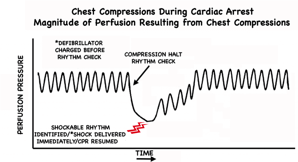
In cardiac arrest care it is well accepted that time to defibrillation is closely correlated with survival and outcome [1]. There has also been a lot of focus over the years on limiting interruptions in chest compressions during CPR. In fact, this concept has become a major focus of the current AHA Guidelines. Why? Because we know interruptions are bad [2,3]. One particular aspect of CPR that has gotten a lot of attention in this regard is the peri-shock period. It has been well established that longer pre- and peri-shock pauses are independently associated with decreased chance of survival [4,5]. Can we do better to shock sooner and minimize these pauses?

Current ACLS standards

Traditionally during manual CPR when a shockable rhythm is encountered at the rhythm check, providers will charge the uncharged defibrillator at that time. In the meantime, chest compressions are typically resumed while waiting for the defibrillator to charge. Once the defibrillator has finished charging, providers are forced to pause yet again in order to “clear” for shock delivery.

“When a rhythm check by a manual defibrillator reveals VF/VT, the first provider should resume CPR while the second provider charges the defibrillator. Once the defibrillator is charged, CPR is paused to ‘clear’ the patient for shock delivery. After the patient is ‘clear,’ the second provider gives a single shock as quickly as possible to minimize the interruption in chest compressions (‘hands-off interval’).” – 2010 AHA/ACC guidelines


Pre-Charge the Defibrillator figure 1

Figure 1: Perfusion pressure changes during CPR without pre-charging the defibrillator. Once a shockable rhythm is identified, there is a delay in shock delivery while awaiting defibrillator charging. There is not just 1 but 2 interruptions in chest compressions. The exact decrease in perfusion pressure during these pauses is variable. Modified from [2].


Trick of the Trade:

Pre-charge the defibrillator during the active chest compression phase of CPR in anticipation of a shockable rhythm

Why should we wait until a shockable rhythm is encountered at the rhythm check point to charge the defibrillator? This makes little sense. Charging the defibrillator prior to the rhythm check is far more logical. With the defibrillator already charged and ready to go, if a shockable rhythm is encountered at the rhythm check point, the shock can be delivered immediately; and importantly, the second interruption is averted entirely.


Pre-Charge the Defibrillator CPR Perfusion Pressure 2

Figure 2: Perfusion pressure changes during CPR with pre-charging the defibrillator. Not only is the shock delivered earlier but the second interruption seen in Figure 1 is avoided completely. Modified from [2].


Logistical tips: How to do this

The most important key to ensuring this process runs smoothly lies in preparation and team briefing prior to patient arrival. There must be clear understanding and communication. The timekeeper is tasked with announcing prior to all rhythm checks (15-30 seconds prior is reasonable) that the rhythm check is approaching. Example:

”In 20 seconds we are due for a rhythm check.”

At this point the team member running the defibrillator charges the defibrillator as chest compressions continue uninterrupted until the rhythm check.

With the defibrillator pre-charged, the team is armed and ready to combat a shockable rhythm immediately as it is encountered at the rhythm check.

What if the rhythm check reveals a non-shockable rhythm? 

If the rhythm check happens to reveal a non-shockable rhythm, CPR can continue as per usual without any alteration. The charge can be manually disarmed, but note that current defibrillators will “hold” the shock for some time (~60 seconds), and if the shock is not delivered in this time frame, the charge will automatically dissipate and require re-charging. (This is why pre-charging should take place within this set time period prior to a rhythm check.) Simply test your particular defibrillator to figure out exactly how long it holds the charge.

Why is pre-charging the defibrillator not in the AHA/ACLS guidelines?

The only arguments I have ever heard against the implementation of this strategy is that it may increase the incidence of inappropriate or inadvertent shocks. Presumably, these theoretical concerns have prevented this technique from being recommended as standard practice.

Are there any studies on pre-charging the defibrillator? 

A multicenter retrospective study was published in 2010 by Edelson et al [6]. Data were gathered from CPR-sensing defibrillator transcripts over a 3-year period. They looked at 680 charge-cycles from 244 cardiac arrests. Charging during compressions correlated with a decrease in median pre-shock pause and total hands-off time in the 30 seconds preceding defibrillation. Interestingly, there was no difference in inappropriate shocks, and there was only one instance of inadvertent shock administration during compressions (which went unnoticed by the compressor).

Discussion

There will likely never be robust data looking at this particular aspect of CPR. Pre-charging the defibrillator is a small thing, but with potentially huge impact. It can be easily taught, easily learned, and is free. I have personally been practicing CPR this way for years now. It has been my experience that it is an incredibly smooth process. All it takes is a little practice and team briefing to ensure all providers are on the same page.

Even with the implementation of other recent techniques that may similarly minimize pauses in chest compressions, such as mechanical CPR and “hands-on defibrillation” (which is of questionable safety), pre-charging the defibrillator still decreases time to defibrillation.

Does it improve outcomes? We may never definitively know. Will it ever make the guidelines? I suspect that eventually it will, perhaps with verbiage similar to something like this:

“It is reasonable to consider pre-charging the defibrillator during chest compressions…”

Until then, it is my opinion that based on early literature, logic, and reasoning, pre-charging the defibrillator in anticipation of a shockable rhythm at the rhythm check is how we should be running our codes.

Conclusion 

Pre-charging the defibrillator during chest compressions in anticipation of a shockable rhythm at the rhythm check shortens time to defibrillation and minimizes the number of pauses in chest compressions during CPR.


References

  1. Chan PS, Krumholz HM, Nichol G, Nallamothu BK, American Heart Association National Registry of Cardiopulmonary Resuscitation Investigators. Delayed time to defibrillation after in-hospital cardiac arrest. N Engl J Med. 2008; 358(1):9-17. PMID: 18172170
  2. Berg RA, Sanders AB, Kern KB, et al. Adverse hemodynamic effects of interrupting chest compressions for rescue breathing during cardiopulmonary resuscitation for ventricular fibrillation cardiac arrest. Circulation. 2001; 104(20):2465-70. PMID: 11705826
  3. Cunningham LM et al. Cardiopulmonary resuscitation for cardiac arrest: the importance of uninterrupted chest compressions in cardiac arrest resuscitation. Am J Emerg Med 2012; 30 (8): 1630 – 8. PMID: 2263371.Clement RA. An extension of Helmholtz’s explanation of Listing’s law. Ophthalmic Physiol Opt. 1990; 10(4):373-80. PMID: 2263371
  4. Brouwer TF, Walker RG, Chapman FW, Koster RW. Association Between Chest Compression Interruptions and Clinical Outcomes of Ventricular Fibrillation Out-of-Hospital Cardiac Arrest. Circulation. 2015; 132(11):1030-7. PMID: 26253757
  5. Cheskes S, Schmicker RH, Christenson J, et al. Perishock pause: an independent predictor of survival from out-of-hospital shockable cardiac arrest. Circulation. 2011; 124(1):58-66. PMID: 21690495
  6. Edelson DP, Robertson-Dick BJ, Yuen TC, et al. Safety and efficacy of defibrillator charging during ongoing chest compressions: a multi-center study. Resuscitation. 2010; 81(11):1521-6. PMID: 20807672

ExpertPeerReviewStamp2x200

Author information

Sam Ghali, MD

Sam Ghali, MD

Clinical Assistant Professor of Emergency Medicine
University of Kentucky

The post Trick of the Trade: Pre-Charge the Defibrillator appeared first on ALiEM.

Managing migraine headaches in complicated patients

$
0
0

migraineCase vignette: A 42-year-old female presents at 10 pm with a throbbing right frontal headache associated with nausea, vomiting, photophobia, and phonophobia. The headache is severe, rated as “10” on a 0 to 10 triage pain scale. The headache began gradually while the patient was at work at 2 pm. Since 2 pm, she has taken 2 tablets of naproxen 500 mg and 2 tablets of sumatriptan 100 mg without relief.

The patient has a diagnosis of migraine without aura. She reports 12 attacks per month. The headache is similar to her previous migraine headaches. She is forced to present to an Emergency Department (ED) on average 2 times per month for management of migraine refractory to oral therapy. She reports a history of dystonic reactions and akathisia after receiving IV dopamine antagonists during a previous ED visit. The physical exam is non-contributory including a normal neurological exam, normal visual fields and fundoscopic exam, and no signs of a head or face infection. When you are done evaluating her, the patient reports that she usually gets relief with 3 doses of hydromorphone 2 mg + diphenhydramine 50 mg IM, and asks that you administer her usual treatment. What do you do?

Background

Migraine is a neurological disorder characterized by recurrent painful headaches and abnormal processing of sensory input resulting in symptoms such as photophobia, phonophobia, and osmophobia [1]. Central to disease pathogenesis is abnormal activation of nociceptive pathways [2]. Disease severity ranges from mild to severe. Patients at one end of the spectrum have rare episodic headaches. On the other end are patients who have headaches on more days than not, patients who are functionally impaired by their headaches, and patients who frequently cannot participate fully in work or social activities. Chronic migraine, a sub-type of migraine defined by ≥15 days with headache for at least 3 consecutive months, is experienced by 1-3% of the general population [3].

ED use for treatment of migraine is common. 1.2 million patients present to U.S. ED’s annually for management of this primary headache disorder [4]. Parenteral opioids are used to treat the acute headache in slightly more than 50% of all ED visits [4]. Multiple authorities have cautioned against the use of opioids for migraine [5,6]. However, the frequent use of opioids has continued unabated, despite the publication in the EM, neurology, and headache literature of dozens of randomized controlled trials (RCTs) demonstrating safety and efficacy of parenteral alternatives, most notably dopamine antagonists and non-steroidal anti-inflammatory drugs [7].

Opioids have been associated with a variety of poor outcomes in migraine patients including:

  1. Progression of the underlying migraine disorder from episodic to chronic migraine [8]
  2. Increased frequency of return visits to ED [9]
  3. Less responsiveness to subsequent treatment with triptans [10]
  4. Less frequent headache relief than patients who received dihydroergotamine or dopamine antagonists [11]

In contrast, a high quality, ED-based RCT did not demonstrate more harm from 1 or 2 doses of meperidine than from dihydroergotamine [12]. Hydromorphone, the parenteral opioid currently used most commonly in U.S. EDs [4], has never been studied experimentally in migraine patients. However, given the wide range of parenteral alternatives, the possibility that opioids may worsen the underlying migraine disorder, and the fact that they are less efficacious than other treatments, opioids should not be offered as first- or second-line therapy for patients who present de novo to an ED with an acute migraine (assuming no contraindications to alternative medications).

Questions:

1) Other than opioids, what parenteral therapies can be offered to this patient?

The 3 classes of parenteral therapeutics with the most evidence supporting safety and efficacy for use as first-line therapy for migraine are the following [13]:

  1. Dopamine antagonists
  2. NSAIDs
  3. Subcutaneous sumatriptan

However, this patient has relative contraindications to each of these. Other parenteral medications used for migraine are listed in the following table.

Table: Alternative parenteral migraine therapies

Agent Dose Adverse events Evidence supporting efficacy Notes
Acetaminophen (APAP) [17, 18] 1 gm IV Well tolerated In one trial, IV APAP did no better than placebo. In another, IV APAP was comparable to an IV NSAID.
Dihydroergotamine [19] 0.5 mg -1 mg IV infusion Nausea is common. Pre-treat with anti-emetics. In one trial, DHE was less effective than sumatriptan at 2 hours but more effective by 4 and 24 hours. Use cautiously in patients with cardiovascular risk factors.
Ketamine [20] 0.08 mg/kg SC Fatigue, delirium In one low quality cross-over RCT, ketamine outperformed placebo.
Magnesium [21-24] 1-2 gm IV Flushing In RCTs of varying quality, IV mg did not consistently outperform placebo Efficacy data is most compelling for migraine with aura.
Octreotide [25] 0.1 mg SC Diarrhea, injection site reactions In a high quality RCT, octreotide did not outperform placebo
Propofol [26,27]

10 mg IV every 10 minutes as needed up to 80 mg

Or

30-40 mg IV with 10-20 mg bolus every 3-5 minutes up to 120 mg

Sedation, hypoxia In a low quality RCT, propofol outperformed dexamethasone. In another low quality trial, propofol outperformed sumatriptan. It is not clear whether the migraine returns after propofol administration has been completed. Previous ALiEM post on migraines and propofol.
Valproic acid [28,29] 1000 mg IV Well tolerated In a high quality RCT, valproate was outperformed by metoclopramide and ketorolac. In a lower quality RCT, valproate was comparable to IV aspirin.

APAP= acetaminophen; DHE= dihydroergotamine; Mg= magnesium


In some patients, greater occipital nerve blocks with a long-acting local anesthetic such as bupivaciane may play a role [14]. While the above alternative parenteral therapies may benefit this patient, available evidence regarding risks and benefits does not dictate that these other therapies must be offered prior to use of opioids.

2) Does the fact that this patient makes frequent use of the ED indicate an unmet medical need?

As with congestive heart failure and asthma, frequent use of an ED for migraine is associated with worse underlying disease [15]. These frequent users are more likely to have chronic migraines (> 15 headache days per month) and psychiatric co-morbidities [15]. Concomitant medication overuse headache, a disorder defined by an upward spiral of increasing headache frequency in the setting of increased usage of analgesic or migraine medication, is also common [16]. Management of complicated patients with migraines is exceedingly difficult, particularly during a busy ED shift, and may lead to frustration for both the healthcare practitioner and the patient. Ideally, outpatient healthcare practitioners with appropriate expertise should direct management of complicated patients with migraines.

3) Should the patient be administered 3 doses of hydromorphone 2 mg + diphenhydramine 50 mg IM as she wishes?

Management of chronic pain patients can be trying and demoralizing for emergency physicians because the underlying problem cannot be solved, and all avenues of treatment are flawed.  Allowing the patient to suffer without appropriate justification is cruel. Delaying opioid administration during good faith efforts to identify alternative effective therapeutic agents is reasonable. Withholding opioids on principle is problematic because for most patients in most circumstances, published data do not establish that the benefit of pain relief is outweighed by the potential for opioid induced harm. On the other hand, thoughtlessly acquiescing to repeated requests for opioids during multiple ED visits is a violation of good medical practice, because of the concern of exacerbating the underlying migraine disorder, which could result in more ED visits, increased number of headache days, and the potential to cause refractoriness to standard migraine medication. One might compare it to administering antibiotics for bronchitis.

Case Resolution

The best solution for the patient in the case vignette is to administer parenteral opioids only as rescue therapy for patients who adhere to an established outpatient plan of care. Acutely, the patient should not be allowed to suffer. However distasteful it may be, the harm arising from 3 isolated doses of parenteral opioids during one ED visit is unlikely to be either long-lasting or severe. But a prerequisite to treatment with opioids during a subsequent visit should be adherence to appropriate outpatient treatment: specifically, patients who require parenteral opioids for migraines should regularly attend outpatient appointments with an appropriate healthcare provider within the ED’s healthcare system.

Department-wide opioid policies are essential, as physician to physician variability in care may undermine a strict approach to opioids. Ideally, a committee with relevant expertise can monitor frequently presenting pain patients and develop patient-specific interventions that will be enforced by all practitioners during subsequent visits. If need be, the terms of treatment can be reinforced with a written document (example in the Appendix). This written document is not meant to be legally binding, but should be used to establish expectations. The last thing a busy emergency physician needs is a battle over opioids with a frequently presenting migraine patient. But before discharge, there should be a conversation about expectations during future ED visits. This will contribute to increased satisfaction for both the provider and the patient.


Top image: (c) Can Stock Photo

REFERENCES
APPENDIX: Sample document to establish expectations for ED patients who require opioids


Author information

Cindy Prettypaul

Cindy Prettypaul

Medical Student
Lake Erie College of Osteopathic Medicine
Seton Hill

The post Managing migraine headaches in complicated patients appeared first on ALiEM.

PEM Pearls: This may hurt! How to manage pediatric anxiety in the ED

$
0
0

screaming child

Pain and anxiety in the emergency department (ED) are two of the most common things we see in children. Pediatric patients, whether first time visitors or those with chronic illnesses, can exhibit marked anxiety and fear when in the ED setting. Child development, parenting styles and prior medical experiences will  guide their reactions in these cases. Practitioners must have a unique set of tools to work with these children and understand the optimal methods for providing care, while decreasing some of these normal reactions to a stressful environment. The most important part of treating anxiety and fear in children is recognizing it early. While pharmacologic interventions can adequately treat pain and anxiety in children, there are quick and effective approaches to avoid these medicines in many cases. Below is a structured approach to assess and reduce anxiety during examination:

The staged evaluation of a child

While a gradual approach to interacting with a child in the ED can take time, the extra 2-4 minutes you spend assessing and interacting with a child may have profound effects on the care following this period. In addition, your approach will be greatly appreciated by the observing parents. So where does the staging start?

  • Vital signs!
    • Are they crying during the blood pressure check or is the child screaming from across the room? These signs will likely indicate a certain type of patient. It also may suggest an opportunity to hold off on immediate evaluation by you, if not emergently required.
  • Outside of the room
    • Where is the child? Are they playing by themselves? Peering out of the door or curtain? A child who may need a more staged approach, may be sitting on or close to the parent.
  • In the room
    • Keep your distance at first and take the “anxiety pulse” of the room. Go in calmly and gradually make movements to engage the child. If they are clinging to the parent, start your interaction with the parent and then engage the child. Toys, bubbles, and other tools can help here. Also, take note of the parental emotions – if they are anxious, working on them may help with the child’s reception.
  • Engagement
    • Aim to stimulate curiosity. Does the child have funny shoes or socks on that you can comment on, or a special stuffed animal with them? Have them help you with a medical tool – place the otoscope cap on or use a mask as a pretend hat. Never start with the painful body area (or the ears!) – start distally and move forward as tolerated.
  • Pitfalls
    • If there is marked pain, treat it! Treating obvious pain promptly, such as with intranasal fentanyl,  will help with your later evaluation, whereas extensively evaluating before pain med administration, may trigger worse anxiety.
    • Language is key: Be cautious of language that creates false expectations, such as telling them that something which will hurt, won’t hurt. Give them options – “Some children say it feels warm, some say it feels cold”, etc.
    • Backpedal: If you step too far over the “anxiety boundary” of the child, backpedal a little bit – re-engage them with a similar technique and go from there. Expect to go back and forth
    • Coach residents and trainees who aren’t experienced before they go into rooms. Don’t expect that young trainees naturally know how to interact with children – appropriately coaching them on these techniques can save you, families, and children lots of headaches!

To learn more, check out Dr. Green-Hopkins, who will be speaking at High Risk Emergency Medicine, June 9-11, 2016 in San Francisco!

Author information

Israel Green-Hopkins, MD

Israel Green-Hopkins, MD

Assistant Clinical Professor
Pediatric Emergency Medicine and Pediatrics
Benioff Children's Hospital
University of California, San Francisco

The post PEM Pearls: This may hurt! How to manage pediatric anxiety in the ED appeared first on ALiEM.

5 Tips for Managing Pain in Older Adults

$
0
0

pain
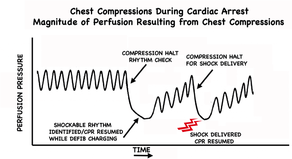
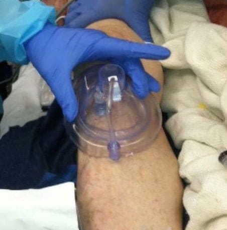
Pain is the most common reason people seek care in Emergency Departments. In addition to diagnosing the cause of the pain, a major goal of emergency physicians (EPs) is to relieve pain. However, medications that treat pain can have their own set of problems and side effects. The risks of treatment are particularly pronounced in older adults, who are often more sensitive to the sedating effects of medications, and are more prone to side effects such as renal failure. EPs frequently have to find the balance between controlling pain and preventing side effects. Untreated pain has large personal, emotional, and financial costs, and more effective, multi-modal pain management can help reduce the burden that acute and chronic pain place on patients [1]. There is evidence that older adults are less likely to receive pain medication in the ED [2,3]. The first step to improving, is being aware of the potential tendency to under-treat pain in older adults. Here are 5 tips to help you effectively manage pain in older adults on your next shift.

1. Know the risks of the medications we use and which patients will be at highest risk.

a) IV opioids. Morphine, hydromorphone, and fentanyl are commonly used in the ED to control moderate to severe pain. They come with the risks of sedation and respiratory depression, particularly when combined with other sedating medications such as benzodiazepines. Older adults, particularly those who have lower body mass and who have more underlying frailty, are at risk for respiratory depression with smaller doses than younger, healthier patients.

b) PO opioids: PO versions are less sedating than IV opioids, and are usually used on discharge for patients who will continue to have pain, such as from a fracture. In older adults they can increase the risk of falls [4] and also cause constipation, which can lead to abdominal pain and additional ED visits.

c) NSAIDs: Older adults are more prone to renal failure, which can be triggered by NSAIDs. This can occur even with a short course of NSAIDs [5]. In terms of long term treatment, GI bleeding and cardiovascular risks are also potential complications. They should certainly be avoided in patients who have known underlying renal failure.

d) Acetaminophen: This is relatively safe in older adults. It is a great go-to, first line med for mild pain, or as an adjunctive for moderate pain.

e) Gabapentin: For neuropathic pain, gabapentin is great, and has few side effects. It can also be used in combination with morphine, and can help reduce the dose of each medication needed while improving pain scores [6].

2. Be attuned to signs of pain, and ask if the patient needs more treatment.

If the patient is able to answer questions then ask if they are in pain and if they want pain medications. A rookie mistake I have made in the past is asking a patient if they are in pain, and then assuming that they want medications. Some patients will opt to not take any medications while the pain is manageable. So asking specifically if they want medication is helpful and saves nursing time so that they do not have to pull the med and then restock it. When reassessing, it also more helpful to ask whether they want more pain medications, rather than to just ask if they still have pain. Most pain medications will not completely remove their pain. The goal is to reduce it to a level that is tolerable, without causing dangerous side effects.

For patients who are nonverbal or who have cognitive impairment, assessing pain can be more challenging than simply asking the patient. Most hospitals use numeric 1-10 pain scales to help assess a patient’s need for medications. However, these scales can fail patients who have cognitive impairments, or have difficulties expressing themselves due to aphasia, prior strokes, or who are acutely ill and delirious. There are scales that have been created to assess pain in patients with cognitive impairment using clues such as patient posturing, facial expressions, negative vocalizations, and vital signs [7]. However, for general practice, if the patient appears uncomfortable, is moaning, has unexplained tachycardia, is grimacing, or curled up in a ball, those can all be signs of pain! Family members and caregivers who are used to the patient’s baseline can also help give input as to whether the patient appears uncomfortable or if their positioning and facial expression is at their baseline. If nonverbal patients appear uncomfortable, it is important to treat their pain. Ongoing pain can contribute to delirium as well as suffering.

3. Start low and go slow.

For patients with mild pain, try non-opioid medications. Try acetaminophen first, or in very healthy patients, you can consider an NSAID. For moderate or severe pain, opioids are appropriate, but consider a lower dose than younger adults, and place patients on a monitor to pick up any respiratory depression or hypoxia. While in younger, healthier adults, a typical starting dose of morphine is 4-8 mg IV, in frail or very elderly adults, 2-4 mg IV may be more appropriate. However it is very important to reassess frequently! If 2 mg of morphine is not providing adequate pain relief 15 minutes after administration, then another 2 mg can be given. This concept also applies to procedural sedation in older adults.

4. Try alternative pain control methods and involve other specialties.

  1. Topicals: Topical NSAIDs are useful for mild joint aches (particularly knee pain) from osteoarthritis, with little systemic absorption. Topical lidocaine, such as a lidocaine patch, is helpful and has a good side-effect profile for musculoskeletal back pain, or for post-herpetic neuralgia.
  2. Regional Anesthesia: Femoral nerve blocks and fascia iliaca compartment blocks are commonly used to reduce pain associated with hip and femoral fractures, and reduce the risks associated with IV opioid administration. Though femoral nerve blocks are the most frequent block used [8], there are many different nerve blocks that can be safely performed in the ED, including blocks or regional anesthesia for joint dislocations, upper extremity and lower extremity fractures, rib fractures and facial or dental injuries or lacerations. These can provide both pain control and anesthesia to allow repair of lacerations or reductions [9-12].
  3. Outpatient referral for physical therapy, or an evaluation by physical medicine and rehab for certain musculoskeletal pains could help the patient long-term, to provide therapy to prevent or help alleviate the pain when it recurs. For patients with chronic pain, a pain clinic referral may help them get the regular care and monitoring they need, and prevent the need for future ED visits.

5. Have a safe discharge plan.

It is important to carefully explain the new medications that you are prescribing, and to write out your instructions for the patient or their caregiver to review later. For example, for a patient with a fracture explain the plan for them to take scheduled acetaminophen and then oxycodone if needed every 8 hours. Avoid using medical terms like “PRN”. Explain that the medications can make them dizzy or sleepy which could put them at risk for falls. Finally, explain the need for Senna and Colace for constipation. Without a good explanation of the discharge plan, patients may simply add their PRN opioid to their scheduled box of medications, and end up taking it even if they don’t need it.

It is also a good idea to have an early follow up plan. If you are able to make an appointment for the patient to see their regular physician, that is best. If not, then encourage them to do so. It is important to have early follow up to reassess the original cause of the pain, as well as the effectiveness of the pain medication, and any side effects that may have arisen.


References

  1. Sinatra R. Causes and consequences of inadequate management of acute pain. Pain Med. 2010;11(12):1859-1871. PMID 21040438
  2. Gilron I, Bailey JM, Tu D, Holden RR, Weaver DF, Houlden RL. Morphine, gabapentin, or their combination for neuropathic pain. N Engl J Med. 2005;352(13):1324-1334. PMID: 15800228
  3. Warden V, Hurley AC, Volicer L. Development and psychometric evaluation of the pain assessment in advanced dementia (PAINAD) scale. J Am Med Dir Assoc. 2003;4(1):9-15. PMID 12807591
  4. Platts-Mills TF, Esserman DA, Brown DL, Bortsov AV, Sloane PD, McLean SA. Older US emergency department patients are less likely to receive pain medication than younger patients: Results from a national survey. Ann Emerg Med. 2012;60(2):199-206. PMID: 22032803
  5. Terrell KM, Hui SL, Castelluccio P, Kroenke K, McGrath RB, Miller DK. Analgesic prescribing for patients who are discharged from an emergency department. Pain Med. 2010;11(7):1072-1077. PMID: 20642733
  6. Rolita L, Spegman A, Tang X, Cronstein BN. Greater number of narcotic analgesic prescriptions for osteoarthritis is associated with falls and fractures in elderly adults. J Am Geriatr Soc. 2013;61(3):335-340. PMID: 23452054
  7. Platts-Mills TF, Richmond NL, Hunold KM, Bowling CB. Life-threatening hyperkalemia after 2 days of ibuprofen. Am J Emerg Med. 2013;31(2):465.e1-465.e2. PMID: 23407039
  8. Amini R, Kartchner JZ, Nagdev A, Adhikari S. Ultrasound-guided nerve blocks in emergency medicine practice. J Ultrasound Med. 2016;35(4):731-736. PMID 26931789
  9. Bhoi S, Chandra A, Galwankar S. Ultrasound-guided nerve blocks in the emergency department. J Emerg Trauma Shock. 2010;3(1):82-88. PMID 20165729
  10. Bhoi S, Sinha TP, Rodha M, Bhasin A, Ramchandani R, Galwankar S. Feasibility and safety of ultrasound-guided nerve block for management of limb injuries by emergency care physicians. J Emerg Trauma Shock. 2012;5(1):28-32. PMID 22416151
  11. Choi JJ, Lin E, Gadsden J. Regional anesthesia for trauma outside the operating theatre. Curr Opin Anaesthesiol. 2013;26(4):495-500. PMID 23673991
  12. Moskovitz JB, Sabatino F. Regional nerve blocks of the face. Emerg Med Clin North Am. 2013;31(2):517-527. PMID 23601486

Image credit [1]

Author information

Christina Shenvi, MD PhD

ALiEM Associate Editor
Assistant Professor
Assistant Residency Director
University of North Carolina
www.gempodcast.com

The post 5 Tips for Managing Pain in Older Adults appeared first on ALiEM.

PV Card: Ankle and Hindfoot Fractures | Quick Reference Guide

$
0
0

Ankle and Hindfoot Fractures Talar Neck Hawkins Classification IVOrthopedic fractures and injuries are commonly managed in the emergency department. Often a quick bedside reference card is needed to remind the clinician about the acute management and follow-up instructions. Thanks to the efforts of a 2015-16 ALiEM Chief Resident Incubator team, we are pleased to announce the first of these reference cards. Ankle and hindfoot fractures covered include ankle malleolar fractures, talus, and calcaneus fractures. These cards were expert reviewed by Dr. Scott Sherman, co-editor of the Emergency Orthopedics textbook (Amazon), and illustrations were created by Dr. Mary Haas.


PV Card: Ankle and Hindfoot Fractures

[Download this PDF]


Ankle Foot Fractures Card final 6-2-16 1

Ankle Foot Fractures Card final 6-2-16 2

Ankle Foot Fractures Card final 6-2-16 3

 [Download this PDF]


Author information

Layla Abubshait, MD

Layla Abubshait, MD

Chief Resident
Emergency Medicine

Central Michigan University

The post PV Card: Ankle and Hindfoot Fractures | Quick Reference Guide appeared first on ALiEM.

Top 5 FOAM Radiology Resources: ALiEM Chief Resident Incubator Recommendations

$
0
0

figure_xray_5391There is no shortage of free open access medical education (FOAM) resources available to the current emergency medicine (EM) learner. It seems that no matter what the concept, FOAM has it covered. And radiology is no different. However, with a specialty as vast as radiology, finding educational material pertinent to the emergency practitioner can be overwhelming. The 2016-2017 ALiEM Chief Resident (CRincubator) class also encountered this when attempting to create an organized FOAM radiology curriculum for EM residents. To tackle this challenge, the chief residents have brought together the best online resources to help EM practitioners gain expertise in the field of radiology.

A collective of over 180 chief residents from across North America were asked to share their favorite radiology sources. Additionally, the internet, literature searches and a review of radiology education pages from major academic institutions were used to gather resources. The compiled material was reviewed by CRincubator collaborators to determine the pros and cons of each resource, the material covered, and how the content may be used in an educational context. Listed below are the sources that best filled a particular niche in radiology education for the EM provider.


Online Text: Radiology Assistant

Editors in Chief: Robin Smithius, MD; Otto van Delden, MD
Website: http://www.radiologyassistant.nl/

Radiology Assistant

Typical page from Radiology Assistant with outline, introduction, and annotated diagram


About Educational site of the Radiological Society of the Netherlands with dedicated sections for each subspecialty of radiology
Pros Designed specifically to provide up-to-date radiological education for radiology residents and radiologists
Great diagrams on anatomy of injuries and definitions of injury type in simplified form
Comprehensive review by type of study and indications for study, which goes beyond learning about particular abnormal findings
Cons May have to wade through radiology-specific discussion (e.g.such as how to protocol a study) to reach EM-salient content


Quick Reference: Radiopaedia

Founder/Editor in Chief: Frank Gaillard, MD
Website: http://radiopaedia.org/

Radiopaedia

Sample article from Radiopaedia about Colles fracture

About Free educational radiology reference resource with one of the web’s largest collections of radiology cases and reference articles
Pros Large repository of images organized by articles and cases (articles in wiki-like form explaining concepts of radiographic findings)
Allows you to research a specific topic or search for thousands of images related to a specific finding or disease process
Most articles elaborate on differential, prognosis, and likely treatment plans
Associated Tumblr and Twitter accounts for interesting cases and radiographic signs
Cons Minimal “clinical correlation” of imaging with management discussion beyond the immediate general disposition of the patient (e.g. operative management or outpatient)


Tutorials/Reference For Beginners: Introduction to Radiology (UVA Health)

Creators/Authors: University of Virginia Health
Website: http://www.med-ed.virginia.edu/courses/rad/

UVA Health

Splenic injury page from UVA Health’s Introduction to Radiology: Emergency Body CT tutorial


About An online, interactive tutorial for introduction to radiology
Pros Self-directed tutorials
Specific tutorials for trauma and emergencies
Expansive
Cons Only tutorials — it does not provide further depth or reference material
Not searchable
Simple hypertext page layout


Smartphone App: Radiology 2.0 – One Night in the ED

Creator: Daniel Cornfeld, MD
Website versioniPhone app link

Radiology 2-0

[LEFT] Blunt chest trauma CT from Radiology 2.0. Touching the screen and moving finger up or down will pan through the different slices. [RIGHT] Blunt chest trauma explanation page

About Introductory educational tool on the CT appearance of basic Emergency Department pathology
Pros Scroll through like a real CT with touchscreen! (The pathology is highlighted later in the explanation page.)
Has sections on blunt abdominal and chest trauma
Thorough reviews of cases with take-home points
Highly rated on the app store for both iPad and iPhone
Free
Cons No neuroradiology or musculoskeletal images
Minimal plain films
Because the text pages do not have images embedded, it reads like a print book


Orthopedic Injuries: Orthobullets

Creator: Derek Moore, MD
Website: http://www.orthobullets.com/

Information page about Supracondylar Fracture from OrthoBullets

Information page about Supracondylar Fracture from OrthoBullets


About Free education site with classic images and management options designed primarily to prepare orthopedic surgeons for standardized exams
Pros Large database of orthopedic injury imaging as well as traumatic neck injuries
Fast, simple bullet points make the site easy to read
Helpful for grading injury type, prognosticating and planning for admission versus outpatient follow-up as well as how to appropriately splint injuries
Cons Simplified
Limited to orthopedics
Mere listing of imaging findings with minimal tutelage about how to interpret the images


Honorable mentions

A large number of educational radiology material are currently available. Some were found to be limited in scope, ease of use, free open access nature or general applicability for the EM physician. While these sources may have limitations they do have features that deserve honorable mention.

  • Lieberman’s eRadiology 
    • Online video lectures/tutorials (some up to 90 minutes in length) similar to a classroom session on topics ranging from MRI to ultrasound, with minimal neuroradiology and limited navigability.
  • Wayne State University Radiology Teaching File
    • Simple hypertext document which is not easily searchable but with a large breadth of pathology based case discussion.
  • HeadNeckBrainSpine
    • Great learning modules on head and neck imaging using CT and MRI and large case library, but limited to neuroradiology.
  • Radiology Masterclass
    • Quick, simple tutorials on a breadth of material (abdomen, chest, and neurological) on a mobile-friendly website, but with minimal distinction between for-pay courses and free tutorials in a busy layout.

Top image: (c) CanStockPhoto

Author information

Christian Rose, MD

Christian Rose, MD

Chief Resident
Emergency Medicine
UCSF-SFGH EM Residency Program

The post Top 5 FOAM Radiology Resources: ALiEM Chief Resident Incubator Recommendations appeared first on ALiEM.

Diagnose on Sight: Swollen Leg

$
0
0

Phlegmasia Cerulea Dolens_1 editedCase: A 58 year-old female presents with a one-day history of worsening right lower extremity pain and swelling, and an acute onset of dyspnea. Her past medical history consists of stage IV renal cell carcinoma diagnosed six months previously. Triage vitals are remarkable for a heart rate of 120 beats per minute and a blood pressure of 68/48 mmHg. What is the diagnosis?


Diagnose on Sight Poll


Answer

Master Clinician Bedside Pearls

Mark Reardon, MD FRCP  
Assistant Professor of Emergency Medicine
University of Ottawa

References

  1. Stallworth JM, Bradham GB, Kletke RR, Price RG, Jr. Phlegmasia Cerulea Dolens: a 10-Year Review. Ann Surg. 1965;161(5): 802-811 PMID: 14290003
  2. Chinsakchai K, Ten Duis K, Moll FL, de Borst GJ. Trends in management of phlegmasia cerulea dolens.Vasc Endovascular Surg. 2011 Jan. 45 (1):5-14 PMID: 21193462
  3. Onuoha CU. Phlegmasia Cerulea Dolens: A Rare Clinical Presentation. Am J Med. 2015 Sep;128(9):e27-8 PMID: 25910785
  4. Kearon C, Akl EA, Comerota AJ et al. Antithrombotic therapy for VTE disease: Antithrombotic Therapy and Prevention of Thrombosis, 9th ed: American College of Chest Physicians Evidence-Based Clinical Practice Guidelines. Chest. 2012 Dec;142(6):1698-1704 PMID: 22315268

Author information

Peter Reardon, MD

Peter Reardon, MD

Emergency Medicine Resident
University of Ottawa

The post Diagnose on Sight: Swollen Leg appeared first on ALiEM.


Ultrasound For The Win! – 63M with an Erythematous Abdomen #US4TW

$
0
0

Welcome to another ultrasound-based case, part of the “Ultrasound For The Win!” (#US4TW) Case Series. In this case series, we focus on a real clinical case where bedside ultrasound changed the management or aided in the diagnosis. In this case, a 63-year-old man presents with a painful, warm, and erythematous area of his abdomen.

Case Presentation

A 63-year-old man with history of diabetes, hypertension, and hyperlipidemia presents with a painful area on his right lower abdomen. He states he noticed pain and redness today, and that it has been worsening over the course of the day. He denies any previous history of similar symptoms and denies trauma. On physical examination, he is a morbidly obese gentleman, in no acute distress. Examination of his abdomen reveals a 10 cm x 12 cm erythematous and tender area on the surface of the right side of his lower abdomen. The area is warm to touch without fluctuance or crepitus. Genitourinary examination is unremarkable.

Vitals

BP 173/82 mmHg
P 111 bpm
RR 23 breaths/min
O2 97% room air
T 37.9 C

Differential Diagnosis

  • Abscess
  • Cellulitis
  • Necrotizing Fasciitis

Laboratory Investigations

  • Total White Blood Cell count: 18 x mm3
  • C-Reactive Protein: 240 mg/L
  • Hemoglobin: 14.3 g/dL
  • Sodium: 139 mmol/L
  • Creatinine: 119 umol/L (or 1.35 mg/dL)
  • Glucose: 12 mmol/L (or 216 mg/dL)
  • Lactate: 4.1 mmol/L

POINT-OF-CARE ULTRASOUND was performed which showed the following:

Figure 1. Cobblestoning of subcutaneous soft tissue with fluid in the deeper fascial plane.
Figure 2. Another view of the cobblestoning of subcutaneous soft tissue and fluid in the deep fascial plane.
Figure 3. Cobblestoning of the subcutaneous tissue (#) and fluid in the deep fascial plane (arrow) is seen.


Ultrasound Image Quality Assurance

The ultrasound images were obtained using the high-frequency linear probe, which is beneficial when attempting to visualize superficial structures within a few centimeters from the surface. The images reveal cobblestoning of the subcutaneous tissue, a non-specific finding that can be seen with cellulitis [Fig. 1, Fig. 2]. Of note, the subcutaneous tissue is uniformly thickened; a comparison of a normal area (e.g. a contralateral limb) can be visualized to confirm abnormal thickening. Deep to the subcutaneous layer is the deep fascial plane, where abnormal fluid is seen in this case [Fig. 3]. These findings can be seen with necrotizing fasciitis. As the disease progresses, abnormal air, visualized as “dirty shadowing” on ultrasound, may be seen in late and more severe cases.

Disposition and Case Conclusion

Given the concerning history and physical examination along with the point-of-care ultrasound concerning for necrotizing fasciitis, empiric antibiotics (IV vancomycin and piperacillin/tazobactam) were given, and surgery was consulted.

The patient was taken to the operating room where a wash out and debridement was performed with a confirmed diagnosis of necrotizing fasciitis. The patient was monitored in the intensive care unit post-operatively and has since been discharged and is doing well.

Background on Necrotizing Fasciitis

Necrotizing fasciitis is rare (with an incidence of 4.3 infections per 100,000 in the United States), but severe soft tissue infection1,2. The most severe form of soft tissue infections, necrotizing fasciitis is a rapidly progressing infection of the subcutaneous tissue and fascia that is potentially limb and life threatening, with a mortality rate of up to 76%2,3. Bacterial enzymes cause tissue necrosis, leading to fluid that can be visualized in the deep fascial layer. The typical bacterial pathogens involved in necrotizing fasciitis include staphylococci, streptococci, and anaerobes, and antibiotic coverage should provide broad coverage for these organisms4. Definitive management requires operative debridement and potential fasciotomy.

The classic physical examination findings of necrotizing fasciitis, including a rapidly progressing area of erythema with ill-defined borders, are often indistinguishable from other soft tissue infections including cellulitis and abscess, especially early in the disease process. Thus, a high index of clinical suspicion is required in the Emergency Department2. While physical exam findings including blistering, hemorrhagic bullae, and crepitus can increase the suspicion of necrotizing fasciitis, these are often late findings seen only in severe and progressed cases2. While necrotizing fasciitis is considered a clinical diagnosis, there may be some utility for laboratory tests and point-of-care ultrasound to aid in risk-stratifying equivocal cases.

LRINEC Score

The LRINEC (Laboratory Risk Indicator for Necrotizing Fasciitis) score utilizes 6 common laboratory tests to risk stratify patients with concern for possible necrotizing fasciitis (Table 1)3. A score of ≥6 should raise your suspicion of the diagnosis, while a score of ≥8 is strongly predictive of necrotizing fasciitis3. In this case, the LRINEC score is 6, which increases the suspicion of necrotizing fasciitis.

Table 1. Laboratory Risk Indicator for Necrotizing Fasciitis (LRINEC) Score

LRINEC Score > 6 should raise suspicion of necrotizing fasciitis. Score > 8 is strongly predictive of necrotizing fasciitis. (Modified from Wong et al.)
Lab, Units Score
C-Reactive Protein, mg/L
< 150 0
≥ 150 4
White cell count, per mm3
< 15 0
15 – 25 1
> 25 2
Hemoglobin, g/dL
> 13.5 0
11 – 13.5 1
< 11 2
Sodium, mmol/L
≥ 135 0
< 135 2
Creatinine
≤ 141 mmol/L or 1.6 mg/dl 0
> 141 mmol/L or 1.6 mg/dL 2
Glucose
≤ 10 mmol/L or 180 mg/dL 0
> 10 mmol/L or 180 mg/dL 1

Ultrasound Findings for Necrotizing Fasciitis

Ultrasound can also be used to identify patients with necrotizing fasciitis. While CT and MRI have been the more traditionally used imaging modalities, they are time consuming, costly, and delay the time to definitive operative management. The ultrasonographic findings of necrotizing fasciitis include diffuse thickening of the subcutaneous tissue when compared to the contralateral side or limb, and a layer of fluid seen more than 4 mm deep along the deep fascial layer1. Using these criteria, ultrasound has been shown to be 88.2% sensitive, 93.3% specific, and 91.9% accurate1. As the disease progresses, air within the fascial layer, seen as “dirty shadowing” may be seen. A useful mnemonic has been described in the literature as the STAFF exam (Subcutaneous Thickening, Air, and Fascial Fluid)2.

Necrotizing fasciitis remains a clinical diagnosis, and concern for the disease requires prompt surgical consultation. While laboratory tests (LRINEC score) and ultrasound are beneficial and can aid in the risk stratification and diagnosis of cases, they should not be used solely to rule out the disease.

Take Home Points

  1. Necrotizing fasciitis is a rare but potentially limb and life threatening infection, requiring a high index of clinical suspicion.
  2. While necrotizing fasciitis is a clinical diagnosis, the LRINEC score and point-of-care ultrasound can aid in the risk-stratification and early diagnosis of the disease.
  3. Ultrasonographic findings suggestive of necrotizing fasciitis include:
    • Fascial and subcutaneous thickening
    • Fluid in the deep fascial layer
    • Subcutaneous air

*Note: All identifying information and certain aspects of the case have been changed to maintain patient confidentiality and protected health information (PHI).

References

1.
Yen Z, Wang H, Ma H, Chen S, Chen W. Ultrasonographic screening of clinically-suspected necrotizing fasciitis. Acad Emerg Med. 2002;9(12):1448-1451.
2.
Castleberg E, Jenson N, Dinh V. Diagnosis of necrotizing faciitis with bedside ultrasound: the STAFF Exam. West J Emerg Med. 2014;15(1):111-113.
3.
Wong C, Khin L, Heng K, Tan K, Low C. The LRINEC (Laboratory Risk Indicator for Necrotizing Fasciitis) score: a tool for distinguishing necrotizing fasciitis from other soft tissue infections. Crit Care Med. 2004;32(7):1535-1541.
4.
Green R, Dafoe D, Raffin T. Necrotizing fasciitis. Chest. 1996;110(1):219-229.

Author information

Jeffrey Shih, MD, RDMS

Jeffrey Shih, MD, RDMS

Director, Emergency Ultrasound Fellowship Program,
The Scarborough Hospital
Lecturer,
University of Toronto
Assistant Editor, Ultrasound for the Win Series,
Academic Life in Emergency Medicine

The post Ultrasound For The Win! – 63M with an Erythematous Abdomen #US4TW appeared first on ALiEM.

Lumbar Puncture on an Anticoagulated Patient in the Emergency Department: Is it safe?

$
0
0

lumbar punctureThe lumbar puncture (LP) procedure is commonly performed in the Emergency Department (ED). While minor complications of LP such as post-procedure headache or back pain occur somewhat regularly, significant complications such as post-procedural spinal hematomas, are rare.1 Despite their low incidence, these spinal hematomas are associated with a significant amount of morbidity for the patient and increased medicolegal risk for the provider.

While post-LP bleeding is usually self-limited and relatively inconsequential, the incidence, severity, and consequence of bleeding may be higher for patients on anticoagulation therapy or anti-platelet agents. To date there has been limited discussion in the Emergency Medicine literature evaluating the potential risk of performing a LP in a patient who is anticoagulated. The following are 5 papers addressing the risks.


Risk for patients taking systemic anticoagulants: The evidence

Risk for patients taking antiplatelet agents: The evidence

Overall the data is limited

To date there are limited high-quality studies examining the risk of post-LP bleeding in anticoagulated patients. Only one study specifically examined post-procedure bleeding after lumbar punctures showing a possible increase in the risk of complications in anticoagulated patients. Despite this potential risk, the overall risk of patients developing a complication that needed an intervention was low. The available data from the inpatient and operating room settings shows a possible association between anticoagulants and post-procedural bleeding but suggests that overall patients are at low risk of developing a significant bleeding complication.7

While there are fundamental differences between the operating room and ED settings, the most comprehensive guidelines regarding spinal puncture originate from the anesthesia literature.

  • Recommendations for anticoagulated patients requiring elective spinal anesthesia include discontinuing anticoagulant use for a duration of twice the half-life of the drug and, when possible, normalizing blood coagulation profiles prior to the procedure.
  • For patients with impaired renal function, who are thought to be at an increased risk of coagulopathy, some sources call for a delay of up to 4 days before performing a spinal puncture.8
  • When invasive spinal procedures are performed in anticoagulated patient, current recommendations call for 24-hours of close post-procedure neurologic monitoring.9

Unfortunately for ED providers, these guidelines suggest the need for caution without fully appreciating the emergent nature and technical nuances of LP performed in the ED. While not directly applicable, ED providers need to balance these conservative recommendations with the overall low rate of significant complications following LP.

The Bottom Line

The exact risk of significant bleeding in anticoagulated patients undergoing an emergent LP is unknown and there is not sufficient evidence to suggest that LP should not be performed in the anticoagulated patient. While the overall rate of significant complication is low, there is a high rate of morbidity in patients who develop post-LP bleeding. Given this risk, providers should make the decision to perform an LP on an anticoagulated patient based on factors such as:

  • Age
  • Comorbidities
  • Spinal pathology
  • Reason for anticoagulation
  • Feasibility of reversing anticoagulation
  • Clinical gestalt of the presenting illness

In addition, the overall utility of the test should be balanced with other considerations such as the possibility of empiric treatment or alternative, non-invasive diagnostic tests.

Providers should involve patients in the medical decision making process both before performing a LP in an anticoagulated patient and when determining the appropriate disposition. While some patients with a high post-procedural risk of bleeding may need to be admitted for further monitoring, other patients with a lower risk may be reasonably discharged with a plan for followup, and specific return instructions.


– Pre-publication peer review performed by Andrew Perron, MD and Emily Vance, PharmD –

1.
Boon J, Abrahams P, Meiring J, Welch T. Lumbar puncture: anatomical review of a clinical skill. Clin Anat. 2004;17(7):544-553.
2.
Ruff R, Dougherty J. Complications of lumbar puncture followed by anticoagulation. Stroke. 1981;12(6):879-881.
3.
Domenicucci M, Ramieri A, Ciappetta P, Delfini R. Nontraumatic acute spinal subdural hematoma: report of five cases and review of the literature. J Neurosurg. 1999;91(1 Suppl):65-73.
4.
Moen V, Dahlgren N, Irestedt L. Severe neurological complications after central neuraxial blockades in Sweden 1990-1999. Anesthesiology. 2004;101(4):950-959.
5.
Horlocker T, Wedel D, Schroeder D, et al. Preoperative antiplatelet therapy does not increase the risk of spinal hematoma associated with regional anesthesia. Anesth Analg. 1995;80(2):303-309.
6.
Horlocker T, Bajwa Z, Ashraf Z, et al. Risk assessment of hemorrhagic complications associated with nonsteroidal antiinflammatory medications in ambulatory pain clinic patients undergoing epidural steroid injection. Anesth Analg. 2002;95(6):1691-7, table of contents.
7.
Benzon H, Wong H, Siddiqui T, Ondra S. Caution in performing epidural injections in patients on several antiplatelet drugs. Anesthesiology. 1999;91(5):1558-1559.
8.
Watanabe M, Qureshi A, Siddiqui F. Periprocedural management of patients on dabigatran etexilate treatment. AJNR Am J Neuroradiol. 2011;32(8):E166.
9.
Horlocker T. Regional anaesthesia in the patient receiving antithrombotic and antiplatelet therapy. Br J Anaesth. 2011;107 Suppl 1:i96-106.

Author information

Matthew DeLaney, MD

Matthew DeLaney, MD

Assistant Professor of Emergency Medicine
Assistant Medical Director
University of Alabama at Birmingham

The post Lumbar Puncture on an Anticoagulated Patient in the Emergency Department: Is it safe? appeared first on ALiEM.

AIR Series: Orthopedics Lower Extremity Module (2016)

$
0
0

ALiEM-AIR-Badge only 200x200Welcome to the Orthopedics Lower Extremity Module! After carefully reviewing all relevant posts from the top 50 sites of the Social Media Index the ALiEM AIR Team is proud to present the highest quality orthopedic lower extremity content. Below we have listed our selection of the 3 highest quality blog posts within the past 12 months (as of March 2016) related to orthopedic lower extremity emergencies, curated and approved for residency training by the AIR Series Board. More specifically in this module, we identified 1 AIRs and 2 Honorable Mentions. We recommend programs give 1 hour (20 minutes per article) of III credit for this module.

AIR Stamp of Approval and Honorable Mentions

In an effort to truly emphasize the highest quality posts, we have two subsets of recommended resources. The AIR stamp of approval will only be given to posts scoring above a strict scoring cut-off of ≥30 points (out of 35 total), based on our scoring instrument. The other subset is for “Honorable Mention” posts. These posts have been flagged by and agreed upon by AIR Board members as worthwhile, accurate, unbiased, and appropriately referenced despite an average score.

Take the quiz at ALiEMU

ALiEMU AIR Orthopedics Lower Extremity block quiz

Interested in taking the quiz for fun or asynchronous (Individualized Interactive Instruction) credit? Please go to the above link. You will need to create a one-time login account if you haven’t already.

Highlighted quality posts

Article TitleAuthorDateLinkTitle
SGEM#138: Hip to be Blocked – Regional Nerve Blocks for Hip and Femoral Neck Fractures

Brandon Ritcey, MD, Chris Bond, MDNovember 29, 2015SGEM: Nerve Blocks for Hip Fx'sAIR
Hip Fractures in Older Adults: An Important Source of Morbidity


Chrinstina Shenvi, MD, PhD
September 15, 2015ALiEM: Hip Fx's in the ElderlyHonorable Mention
Episode 40 - Femoral Nerve Blocks & Compartment Syndrome
Unknown AuthorDecember 23, 2015FOAMcast: Fem Nerve Blocks Honorable Mention


If you have any questions or comments, please contact us!


Author information

Andrew Grock, MD

Andrew Grock, MD

Lead Editor/Co-Founder of ALiEM Approved Instructional Resources (AIR)
Assistant Professor of Emergency Medicine
UCLA Emergency Medicine Department

The post AIR Series: Orthopedics Lower Extremity Module (2016) appeared first on ALiEM.

AIR Series: Orthopedics Upper Extremity Module (2016)

$
0
0

air seriesWelcome to the Orthopedics Upper Extremity Module! After carefully reviewing all relevant posts from the top 50 sites of the Social Media Index the ALiEM AIR Team is proud to present the highest quality orthopedic upper extremity content. Below we have listed our selection of the 14 highest quality blog posts within the past 12 months (as of March 2016) related to orthopedic upper extremity emergencies, curated and approved for residency training by the AIR Series Board. More specifically in this module, we identified 2 AIRs and 12 Honorable Mentions. We recommend programs give 5 hours (just over 20 minutes per article) of III credit for this module.


AIR Stamp of Approval and Honorable Mentions

In an effort to truly emphasize the highest quality posts, we have two subsets of recommended resources. The AIR stamp of approval will only be given to posts scoring above a strict scoring cut-off of ≥30 points (out of 35 total), based on our scoring instrument. The other subset is for “Honorable Mention” posts. These posts have been flagged by and agreed upon by AIR Board members as worthwhile, accurate, unbiased, and appropriately referenced despite an average score.

Take the quiz at ALiEMU

ALiEMU AIR Orthopedics Upper Extremity block quiz

Interested in taking the quiz for fun or asynchronous (Individualized Interactive Instruction) credit? Please go to the above link. You will need to create a one-time login account if you haven’t already.


Highlighted quality posts

Article TitleAuthorDateLinkTitle
More Dogma: Epinephrine in Digital Nerve Blocks Rob Bryant, MDSeptember 3, 2015Rebel EM: Epi in DigitsApproved Instructional Resource
A St.Emlyn’s fascia iliaca block updateAlan Grayson , MDJanuary 22, 2016St Emlyn's: Iliaca BlockApproved Instructional Resource
Septic Joint: Reminders, Updates and Pitfalls Matthew A. Heimann, MD and Kevin Barlotta, MD May 9, 2015EMDocs: Septic JointHonorable Mention
Open FracturesRichard B. Moleno, DO, MS and Michael Venezia, DO, MPH December 31, 2015EM Docs: Open FracturesHonorable Mention
Boring Question: Does this pediatric patient require a hard cast ? Patrick Bafuma, PA March 23, 2015CanadiEM: Peds and Hard CastsHonorable Mention
PV Card: Adult scaphoid fracture Sean Kivlehan, MD, MPHFebruary 1, 2016ALiEM: Scaphoid FractureHonorable Mention
Episode 52: Commonly Missed Uncommon Orthopedic InjuriesAnton Helman, MDOctober 1, 2014EM Cases: Missed Ortho InjuriesHonorable Mention
Clinical Question: How effective is intra-articular lidocaine for shoulder reduction? Patrick Bafuma, PAMarch 22, 2016Candi EM: IA LidoHonorable Mention
Handy Knowledge: Subtle and High-Risk Hand Injuries Ryan Mason, MD and Alex St. John, MD September 19, 2015EM Docs: Hand InjuriesHonorable Mention
Episode 58: Tendons and Ligaments – Commonly Missed Uncommon Orthopedic Injuries Part 2 Anton Helman, MDJanuary 1, 2015EM Cases: Missed Ortho Injuries 2Honorable Mention
The Crashing Patient with Long Bone Fractures: A Case of Fat Embolism Syndrome Molly Tollins, MD, Nick Johnson, MDJanuary 21, 2016EM Docs: Fat Embolism 1Honorable Mention
Wrist and Distal Forearm Injuries: Pearls and Pitfalls Jason Brown, MD November 7, 2015EM Docs: Wrist/Forearm InjuriesHonorable Mention
Septic Arthitis Sean Fox, MDAugust 28, 2015PEM Morsels: Septic JointHonorable Mention
Shoulder Dislocation Sean Fox, MDJuly 31, 2015PEM Morsels: Shoulder dislocationHonorable Mention


If you have any questions or comments, please contact us!

Author information

Andrew Grock, MD

Andrew Grock, MD

Lead Editor/Co-Founder of ALiEM Approved Instructional Resources (AIR)
Assistant Professor of Emergency Medicine
UCLA Emergency Medicine Department

The post AIR Series: Orthopedics Upper Extremity Module (2016) appeared first on ALiEM.

AIR Series and AIR-Pro Series are moving to ALiEMU!

$
0
0

AliEMU-Favicon-Turqouise-textThe ALiEM Approved Instructional Resources (AIR) and AIR-Pro series are moving from this ALiEM blog, which uses embedded Google Forms for quizzes into our custom learning management system called ALiEMU. ALiEMU will be our one-stop system for asynchronous learning. For U.S. EM residency programs, this will also serve as a central repository for Individualized Interactive Instruction (III) resources for asynchronous conference credit. The cornerstone e-course already on ALiEMU is CAPSULES — a comprehensive EM pharmacology curriculum, whose authorship and editorial team is led by Dr. Bryan Hayes.

For the AIR and AIR-Pro series, we have over 80 U.S. EM residency programs, 4 international EM programs, and 1 PA program using either or both of these series as a part of their didactic curriculum. With over one year’s worth of educational content, we now want to make the user experience more friendly such that one can longitudinally track progress, and programs and easily report their residents’ total III hours for the academic year to the Residency Review Committee (RRC).

What is Capsules?

Thanks to the ALiEM Capsules Series [background information about the series], which was the premiere longitudinal course to launch ALiEMU in 2015, we have worked out many of the kinks and start-up problems of new platforms. The Capsules series, led by Dr. Bryan Hayes, features an all-star team of pharmacists and EM faculty authoring key chapters and quizzes on all things related to EM pharmacology.

Capsules Screenshot

Create an account on ALiEMU

ALiEMU Register

  • Go to the AIR or AIR-Pro home page.
  • View the desired course module and take the corresponding quizzes for course completion.

AIR sample module

Detailed guides on navigating ALiEMU

Specific information for the PD or APD

  • We now have 3 longitudinal series available for III credit: AIR, AIR-Pro, and Capsules
  • Request “Educator Dashboard” access by clicking on the checkbox when registering. Only the following people will be granted access:
    • PDs
    • APDs
    • Program coordinators
    • PD-approved administrative assistants
    • PD-approved Chief Residents who specifically working on resident remediation  will be granted access.
  • As your residents complete the AIR, AIR-Pro, and Capsules modules, your Educator Dashboard will be updated in real-time. Your Dashboard allows you to view your entire program’s progress on the website. This can be filtered by DATE or by SERIES for a more limited view.
  • Tracking III hours: For a quick overview and searching for a particular person or timeframe, it is easiest to use the Educator Dashboard. To count and report III hours, it is best to export the CSV file to sort and collate the data to your program’s needs.


Date Filter


Series-Block filter


Frequently asked questions about the AIR and AIR-Pro Series moving to ALiEMU

  1. As a resident, if I completed an AIR or AIR-Pro module before today (July 1, 2016) using the Google Forms quiz, how can my (A)PD access my data?
    • Your (A)PD still access to the master Google spreadsheet.
    • Contact us, if you are an (A)PD and need your login.
  2. How do I contact someone if I encounter a bug or need help with ALiEMU issues?
  3. If am a resident, can I request Educator Dashboard access?
    • Because the Dashboard has data of your fellow residents, we are currently not granting access, unless your PD specifically requests that we grant you access.
  4. Can I get a certificate for completing each AIR or AIR-Pro course?
    • At the end of each course, you are given the option to print or download your certificate (PDF).

Certificate


Acknowledgements

A huge thank-you and virtual standing ovation for the ALiEMU development team of Derek Sifford (Chief Technical Officer), Dr. Chris Gaafary (Chief of Development and Design) and Dr. Jonathan Bronner (Education Design Officer) who worked tirelessly for weeks to make ALiEMU and the AIR/AIR-Pro Series transition a reality. Watching them work and collaborate virtually at a startup-like pace was 99% inspiring and 1% frightening to witness the power of digital technologies.

ALiEMU-Core-team

Also thank-you to Dr. Andy Grock and Dr. Fareen Zaver for facilitating the move of their AIR Series and AIR-Pro Series, respectively. And lastly, thank you to the Council of EM Residency Directors for funding our efforts to create the custom Educator Dashboard for enhanced III tracking.


The ALiEMU Team Members

You can see our entire 50+ member team on ALiEMU’s Our Teams page.

Author information

Michelle Lin, MD

ALiEM Editor-in-Chief
Academy Endowed Chair of EM Education
Professor of Clinical Emergency Medicine
University of California, San Francisco

The post AIR Series and AIR-Pro Series are moving to ALiEMU! appeared first on ALiEM.

Viewing all 273 articles
Browse latest View live


Latest Images