Quantcast
Channel: Clinical – ALiEM
Viewing all 273 articles
Browse latest View live

AIR-Pro Series: Pediatrics (2016)

$
0
0

Below we have listed our selection of the 14 highest quality blog posts related to 5 advanced level questions on pediatric topics posed, curated, and approved for residency training by the AIR-Pro Series Board. The blogs relate to theALiEM-AIR-Badge-PRO-only sm following questions:

  1. Pediatric arrhythmias
  2. Procedural sedation in pediatrics
  3. The neonate in distress
  4. Toddlers with a limp
  5. Pediatric syncope

In this module, we have 10 AIR-Pro’s and 4 honorable mentions. To strive for comprehensiveness, we selected from a broad spectrum of blogs identified through FOAMSearch.net.

This module we also had two editorial board guests trained in Pediatric Emergency Medicine to increase the strength of our recommendations – Dr. Robert Cloutier and Dr. Jason Woods.

AIR-Pro Stamp of Approval and Honorable Mentions

In an effort to truly emphasize the highest quality posts, we have 2 subsets of recommended resources. The AIR-Pro stamp of approval will only be given to posts scoring above a strict scoring cut-off of ≥28 points (out of 35 total), based on our AIR-Pro scoring instrument, which is slightly different from our original AIR Series scoring instrument. The other subset is for “Honorable Mention” posts. These posts have been flagged by and agreed upon by AIR-Pro Board members as worthwhile, accurate, unbiased and useful to senior residents. Only the posts with the AIR-Pro stamp of approval will be part of the quiz needed to obtain III credit. To decrease the repetitive nature of posts relating to these advanced concepts, we did not always include every post found that met the score of ≥28 points.

After reading, please take the quiz. You do not have to complete the honorable mentions to complete the quiz. Feel free to ask questions in the blog comment section below. The AIR Board faculty will answer them within 48 hours of posting. Be sure to include your email or contact information where requested in the Disqus blog comment area, so that you will be notified when we reply.

Optional Background Reading

The following table contains links to basic/background knowledge that may be helpful to review prior to reading these more advanced-level blog posts. These are not part of the III quiz.

Article TitleAuthorDateLink
ECG BasicsSonny TatAug 13, 2014PEM Academy: ECG Basics
ECG CentralMay 2, 2014Don't forget the bubbles: ECG Central
Patwari Academy videos: The Crashing NeonateMichelle LinJuly 7, 2013Patwari Academy videos: The Crashing Neonate

Pediatric Module 2016: Recommended III credit hours

3.5 hours (20 minutes per article, 30 minutes for articles with podcasts)

Article TitleAuthorDateLinkTitle
ECGs: Long QT and BrugadaShilpa Patel & Anne GreenSept 18, 2014PEM Academy: ECGs - Long QT and BrugadaAir Pro
ECGs: Heart block and Sick SinusSonny TatJuly 7, 2014PEM Academy: ECGs - Heart block and Sick SinusAir Pro
Ketamine for AnalgesiaSean FoxApril 11, 2014Pediatric EM Morsels: KetamineAir Pro
KetamineBrad SobolewskiJuly 24, 2013PEM Currents: KetamineAir Pro
The Sick NeonateZach RadwineAug 12, 2014EMDocs: The Sick NeonateAir Pro
A Knackered NeonateChristopher Partyka
July 19, 2013Blunt Dissection: A Knackered NeonateAir Pro
Pediatric LimpRob OrmanFeb 2, 2010ER Cast: Pediatric LimpAir Pro
Toddler's FractureSean FoxFeb 1, 2013Pediatric EM Morsels: Toddler's FractureAir Pro
Brugada in ChildrenSean FoxSept 11, 2015Pediatric EM Morsels: BrugadaAir Pro
Syncope Sunday 4: Syncope ECGsElayne ForbesMarch 15, 2015Dont forget the bubbles: Syncope ECGsAir Pro
The Toxic NeonateRob OrmanMay 26, 2010ER Cast: The Toxic NeonateHonorable Mention
Episode 35: Pediatric Orthopedics Pearls and PitfallsSanjay Mehta & Jonathen PirieOct, 2014Emergency Medicine Cases: Pediatric OrthopedicsHonorable Mention
Pediatric Syncope Zach RadwineJuly 7, 2014EM Docs: Pediatric SyncopeHonorable Mention
Hypertrophic Cardiomyopathy (HCM)Ed Burns, Chris Nickson & Amal MattuJuly 30, 2012Life in the Fast Lane: Hypertrophic Cardiomyopathy Honorable Mention

red_pencil_horizontal_pc_400_clr_3248

Take the quiz below, or click HERE to take you to the quiz site.
Be sure to enter your name and program, if you desire III credit.

Background Information About the AIR-Pro Series

The ALiEM Approved Instructional Resources (AIR)-Pro Series is an effort to reward our residents for the reading and learning they are already doing online. We have created an Individual Interactive Instruction (III) opportunity utilizing Free Open Access Meducation (FOAM) resources for U.S. Emergency Medicine residents. For each module, the AIR-Pro Board curates and scores a list of blogs and podcasts specifically answering questions tailored to the senior resident. A quiz is available to complete after each module to obtain residency conference credit. Once completed, your name and institution will be logged into our private Google Drive database, which participating residency program directors can access to provide proof of completion.

Do you belong to a residency program that is not currently participating? No problem! Any one can read the AIR-Pro series curated post and complete the quiz for educational value!

If a residency program is interested in participating, please contact us!

Author information

Fareen Zaver, MD

Fareen Zaver, MD

Chief Resident
Department of Emergency Medicine
George Washington University

The post AIR-Pro Series: Pediatrics (2016) appeared first on ALiEM.


PV Card: Adult scaphoid fracture

$
0
0

snuffbox tendernessWhat is the most commonly fractured carpal bone in adults? It’s the scaphoid bone. As a bonus it has the dreaded complication of avascular necrosis. So how good are the physical exam and imaging modalities in diagnosing a fracture? What is the likelihood ratio (LR) that snuffbox tenderness predicts a fracture? Bottom lines: The exam is highly sensitive but poorly specific, such that one can only confidently state that a NON-tender snuffbox and scaphoid tubercle essentially rule out an acute scaphoid fracture. Also negative x-rays for patients with scaphoid tenderness still yield a fracture post-test probability of 25%. This PV card breaks down all the LRs [1].

PV Card: Scaphoid Fracture

Scaphoid Fracture PV

You can download this PV card: [MS Word] [PDF].
See other Paucis Verbis cards.

Reference

  1. Carpenter CR, Pines JM, Schuur JD, Muir M, Calfee RP, Raja AS. Adult scaphoid fracture. Acad Emerg Med. 2014; 21(2): 101-21. PMID: 24673666


Author information

Sean Kivlehan, MD MPH

Sean Kivlehan, MD MPH

International Emergency Medicine Fellow

Department of Emergency Medicine
Brigham and Women's Hospital

The post PV Card: Adult scaphoid fracture appeared first on ALiEM.

Must-Know EM Pharmacotherapy Articles of 2015

$
0
0

Top10There is so much literature to sift through each year, it becomes nearly impossible to stay abreast of it. Here is a quick summary of the 10 must-know Emergency Medicine pharmacotherapy articles from 2015, in my humble opinion.


Top 10 EM Pharmacotherapy Articles of 2015

1. IV Magnesium for Acute Migraine Headache2. Ketamine for Alcohol Withdrawal3. Vancomycin Loading in Obese Patients4. Early Glargine Administration at Start of DKA Treatment5. Reversing Dabigatran with Idarucizumab6. Blood Glucose Response to Rescue Dextrose7. Ketamine vs. Morphine for Analgesia in the ED8. Avoid Opioids for Low Back Pain9. Andexanet Alfa for the Reversal of Factor Xa Inhibitor Activity10. Therapeutic Tramadol Use Significantly Increases Seizure RiskBonus ArticleFavorite Article of the Year

Does IV magnesium have a role in the management of acute migraine headache in the ED? A new study says yes. [1]

Intervention

  • 35 patients received IV magnesium 1 gm over 15 minutes.
  • 35 patients received IV dexamethasone 8 mg + IV metoclopramide 10 mg over 15 minutes.
  • Each group contained men and women.
  • Initial pain score 8.2 in dexamethasone/metoclopramide group vs. 8.0 in magnesium group.

What They Found

Magnesium sulfate was more effective in decreasing pain severity at 20-min (pain scale 5.2 vs. 7.4) and 1-h (2.3 vs. 6.0) and 2-h (1.3 vs. 2.5) intervals after treatment (p < 0.0001) compared to treatment with dexamethasone/metoclopramide.

Application to Clinical Practice

Two previous studies found mixed results using magnesium (Corbo 2001, Cete 2005). This new study found that IV magnesium may be an additional option. The authors didn’t compare magnesium to more common treatments such as prochlorperazine or metoclopramide 20 mg (+/- ketorolac and diphenhydramine), which may limit its generalizability. However, magnesium’s pain lowering effect was good regardless of comparator group.

Shahrami A, et al. Comparison of therapeutic effects of magnesium sulfate vs. dexamethasone/metoclopramide on alleviating acute migraine headache. J Emerg Med 2015;48(1):69-76. [PMID 25278139]

Originally posted Jan 3, 2015 as University of Maryland (UMEM, @UMEmergencyMed) pearl.

Background

In addition to the down regulation of GABA receptors in chronic ethanol users, there is an upregulation in NMDA receptor subtypes. Although the pathophysiology is much more complex, when ethanol abstinence occurs, there is a shortage of GABA-mediated CNS inhibition and a surplus of glutamate-mediated CNS excitation. If GABA agonists are the mainstay of treatment, why not also target the NMDA receptor? Enter ketamine.

The Data

Only one study exists and was published recently.

  • Retrospective review of 23 adult patients administered ketamine specifically for management of AWS.
  • Mean time to initiation of ketamine from first treatment of AWS, and total duration of therapy were 33.6 and 55.8 hours, respectively.
  • Mean initial infusion dose and median total infusion rate were 0.21 and 0.20 mg/kg/h, respectively.
  • No change in sedation or alcohol withdrawal scores within 6 hours of ketamine initiation.
  • Median change in benzodiazepine requirements at 12 and 24 hours post-ketamine initiation were -40.0 and -13.3 mg, respectively.
  • One documented adverse reaction of oversedation, requiring dose reduction.
  • Authors concluded that ketamine appears to reduce benzodiazepine requirements and is well tolerated at low doses.

Application to Clinical Practice

While the dexmedetomidine studies should not be using reduction in benzodiazepine requirements as an endpoint, it may be acceptable for ketamine since it actually works on the underlying pathophysiology. More studies are needed but it’s good to see we’re starting to look at it.

Wong A, et al. Evaluation of adjunctive ketamine to benzodiazepines for management of alcohol withdrawal syndrome. Ann Pharmacother 2015;49(1):14-9. [PMID 25325907]

Originally posted May 7, 2015 as UMEM pearl.

Vancomycin guidelines recommend an initial dose of 15-20 mg/kg based on actual body weight (25-30 mg/kg in critically ill patients) (Ryback 2009). The MRSA guidelines further recommend a max dose of 2 gm (Liu 2011).

But, what dose do you give for an obese patient that would require more than 2 gm?

A new study provides some answers to this question. Obese-specific, divided-load dosing achieved trough concentrations of 10 to 20 g/mL for 89% of obese patients within 12 hours of initial dosing and 97% of obese patients within 24 hours of initial dosing.

Application to Clinical Practice

  1. Calculate the total loading dose. At my institution we use actual body weight (the study used IBW).
  2. Divide the total dose to be given every 6 hours until load is complete. We cap each individual dose at 2 gm (the study used 1.5 gm).
  3. Measure a trough level before the third dose.
  4. Change to dosing frequency dictated by renal function once level moves into target range.

Caveats

The study used some more specific dosing calculations based on renal function and percentage above IBW. If patient’s renal function is abnormal, consultation with a pharmacist is recommended.

Denetclaw TH, et al. Performance of a divided-load vancomycin dosing strategy for obese patients. Ann Pharmacother 2015;49(8):861-8. [PMID 2598600]

Originally posted June 6, 2015 as UMEM pearl.

Transitioning Diabetic Ketoacidosis (DKA) patients off an insulin infusion can be challenging. If a long-acting insulin, such as glargine or levemir, is not administered at the correct time to provide extended coverage, patients can revert back into DKA.

Pilot Study

A prospective, randomized, controlled pilot study in 40 patients evaluated administration of glargine within 2 hours of insulin infusion initiation compared to waiting until the anion gap (AG) had closed.

What they did

  • All patients received IV insulin.
  • Experimental: Subcutaneous insulin glargine given within 2 hours of diagnosis.
  • Control: Patients subsequently transitioned to long-acting insulin upon closure of AG.

What they found

Mean time to closure of AG, mean hospital LOS, incidents of hypoglycemia, rates of ICU admission, and ICU LOS were all similar between the groups.

Application to Clinical Practice

Although just a pilot study (using a convenience sample), early glargine administration seemed to be absorbed adequately (based on time to AG closure) and was not associated with increased risk of hypoglycemia. If confirmed in a larger study, this technique could help optimize care of DKA patients in the ED by eliminating the often-mismanaged transition step later on.

Doshi P, et al. Prospective randomized trial of insulin glargine in acute management of diabetic ketoacidosis in the emergency department: a pilot study. Acad Emerg Med 2015;22(6):657-62. [PMID 26013711]

Originally posted July 4, 2015 as UMEM pearl.

The New England Journal of Medicine and Lancet both published studies evaluating idarucizumab for reversal of dabigatran. It is amonoclonal antibody fragment that binds dabigatran with high affinity. Dr. Ryan Radecki summarizes the two articles on his EM Lit of Note blog.

Here are a few take home points from these early studies:

  1. Both studies were funded by Boehringer Ingelheim, who not suprisingly also markets dabigatran. Skepticism is always welcome when the same company makes the drug and the antidote.
  2. The Lancet study was conducted in healthy volunteers, while the NEJM study was conducted in patients needing reversal but lacked a control group.
  3. Idarucizumab seems to reverse laboratory markers of anticoagulation from dabigatran rapidly and completely, including dilute thrombin time and ecarin clotting time. Not all institutions have these assays available.
  4. The dose that seems to ‘work’ the best is 5 gm given IV (two-2.5 gm infusions given no more than 15 minutes apart).
  5. Median investigator-reported time to cessation of bleeding was 11.4 hours in the NEJM study.
  6. 21 of the 90 patients in the NEJM study had ‘serious adverse effects’ including thrombotic events.
  7. The acquisition cost of this medication will most assuredly be high if and when it is FDA-approved in the U.S.

Pollack CV, et al. Idarucizumab for dabigatran reversal. N Engl J Med 2015;373(6):511-20. [PMID 26095746]

Glund S, et al. Safety, tolerability, and efficacy of idarucizumab for the reversal of the anticoagulant effect of dabigatran in health male volunteers: a randomised, placebo-controlled, double-blind phase 1 trial. Lancet 2015;386:680-90. [PMID 26088268]

Originally posted July 9, 2015 as UMEM pearl.

How much does the blood glucose concentration increase when dextrose 50% (D50) is administered?

A new study found a median increase of 4 mg/dL (0.2 mmol/L) per gram of D50 administered.

This retrospective study was conducted in critically ill patients who experienced hypoglycemia while receiving an insulin infusion. While it may not directly apply to all Emergency Department patients, an estimation of the expected blood glucose increase from rescue dextrose is helpful. If the blood glucose doesn’t respond as anticipated, it can help us troubleshoot possible issues (eg, line access).

Murthy MS, et al. Blood glucose response to rescue dextrose in hypoglycemic, critically ill patients receiving an insulin infusion. Ann Pharmacother 2015;49(8):892-6. [PMID 25986006]

Originally posted August 1, 2015 as UMEM pearl.

A new prospective, randomized, double-blind trial compared subdissociative ketamine to morphine for acute pain in the ED.

What they did

  • 45 patients received IV ketamine 0.3 mg/kg (mean baseline pain score 8.6)
  • 45 patients received IV morphine 0.1 mg/kg (mean baseline pain score 8.5)
  • Source of pain was abdominal for ~70% in each group
  • Exclusion criteria was pretty standard

What they found

  • Pain score at 30 minutes: 4.1 for ketamine vs. 3.9 for morphine (p = 0.97)
  • No difference in the incidence of rescue fentanyl analgesia at 30 or 60 minutes
  • No serious adverse events occurred in either group
  • Patients in the ketamine group reported increased minor adverse effects at 15 minutes post-drug administration

Application to clinical practice

  1. In an effort to reduce opioid use in the ED, low-dose ketamine may be a reasonable alternative to opioids for acute analgesia.
  2. State nursing regulations govern who can administer IV ketamine in the ED.
  3. What to prescribe on discharge? Lead author Dr. Motov recommends a “pain syndrome targeted” approach with “patient-specific opioid and non-opioid analgesics.”

Motov S, et al. Intravenous subdissociative-dose ketamine versus morphine for analgesia in the emergency department: a randomized controlled trial. Ann Emerg Med 2015;66:222-9. [PMID 25817884]

If there weren’t enough reasons to avoid opioids, here is another: opioids don’t work for low back pain (LBP).

Objective

A well-done, double-blind, randomized controlled trial from JAMA set out to compare functional outcomes and pain at 1 week and 3 months after an ED visit for acute LBP among patients randomized to a 10-day course of (1) naproxen + placebo; (2) naproxen + cyclobenzaprine; or (3) naproxen + oxycodone/acetaminophen.

Intervention

  • Nontraumatic, nonradicular LBP of 2 weeks’ duration or less
  • All patients were given 20 tablets of naproxen, 500 mg, to be taken twice a day.
    • They were randomized to receive either 60 tablets of placebo; cyclobenzaprine, 5 mg; or oxycodone, 5 mg/acetaminophen, 325 mg. Participants were instructed to take 1 or 2 of these tablets every 8 hours, as needed for LBP.
  • Patients received a standardized 10-minute LBP educational session prior to discharge.

Outcome

Neither oxycodone/acetaminophen nor cyclobenzaprine improved pain or functional outcomes at 1 week compared to placebo, and more adverse effects were noted.

Application to Clinical Practice

Among patients with acute, nontraumatic, nonradicular LBP presenting to the ED, avoid adding opioids or cyclobenzaprine to the standard NSAID therapy.

Friedman BW, et al. Naproxen with Cyclobenzaprine, Oxycodone/Aceaminophen, or Placebo for Treating Acute Low Back Pain: A Randomized Clinical Trial. JAMA 2015;314(15):1572-80. [PMID 26501533]

Not to be outdone by the recent FDA approval of Idarucizumab to reverse dabigatran, a new factor Xa reversal agent is under investigation. “Andexanet binds and sequesters factor Xa inhibitors within the vascular space, thereby restoring the activity of endogenous factor Xa and reducing levels of anticoagulant activity, as assessed by measurement of thrombin generation and anti factor Xa activity, the latter of which is a direct measure of the anticoagulant activity.”

Design

Two parallel randomized, placebo-controlled trials (ANNEXA-A [apixaban] and ANNEXA-R [rivaroxaban]) were conducted in healthy vounteers to evaluate the ability of andexanet to reverse anticoagulation, as measured by the percent change in anti factor Xa activity after administration.

What they Found

Compared to placebo, andexanet significantly reduced anti-factor Xa activity, increased thrombin generation, and decreased unbound drug concentration in both the apixaban and rivaroxaban groups.

Application to Clinical Practice

  1. This drug is not yet FDA approved.
  2. These trials were funded by the maker of andexanet (Portola Pharmaceuticals) and supported by the makers of apixaban and rivaroxaban.
  3. Studies are needed in patients requiring urgent reversal.
  4. The trials looked only at laboratory markers of anticoagulation. We don’t know how fast (or the extent of) the reversal activity is in the clinical setting.

Siegal DM, et al. Andexanet Alfa for the Reversal of Factor Xa Inhibitor Activity. N Engl J Med 2015;373(25)2413-25. [PMID 26559317]

Tramadol has a reputation for being a safe, non-opioid alternative to opioids. Nothing could be further from the truth. Several blogs have published about the dangers of tramadol:

But what about seizure risk? Previous studies have been unable to confirm an increased seizure risk with therapeutic doses of tramadol (Seizure Risk Associated with Tramadol Use from EM PharmD blog). However, a new study refutes that premise.
22% of first-seizure patients had recent tramadol use!

  1. Mean total tramadol dose in last 24 hours (reported): 140 mg
  2. Duration of tramadol use less than 10 days: 84.5%
  3. Seizure within 6 hours of tramadol consumption: 74%

This was a retrospective study without laboratory confirmation of tramadol intake. Nevertheless, it behooves us not to think of tramadol as a safer alternative to opioids. It is an opioid after all, and it comes with significant adverse effects.

Asadi P, et al. Prevalence of Tramadol Consumption in First Seizure patients; a One-Year Cross-sectional Study. Emerg (Tehran) 2015;3(4):159-61. [PMID 26495407]

Pickard R, et al. Medical expulsive therapy  in adults with ureteric colic: a multicentre, randomised, placebo-controlled trial. Lancet 2015;386:341-9. [PMID 25998582]

Take Home: Tamsulosin 0.4 mg and nifedipine 30 mg are not effective at decreasing the need for further treatment to achieve stone clearance in 4 weeks for patients with expectantly managed ureteric colic.

It’s not technically a pharmacotherapy article, but this is my favorite article of the year:

Favaloro EJ, et al. Laboratory Testing in the Era of Direct or Non–Vitamin K Antagonist Oral Anticoagulants: A Practical Guide to Measuring Their Activity and Avoiding Diagnostic Errors. Semin Thromb Hemost 2015;41:208–27. [PMID 25703514]

It’s the most comprehensive and practical review of laboratory interpretation for oral anticoagulants I’ve seen. It includes excellent algorithms for how to confirm/exclude various oral anticoagulants based on commonly available labs. An absolute must-read and an outstanding resource to keep in your Dropbox or Google Drive.

Author information

Bryan D. Hayes, PharmD, FAACT

Bryan D. Hayes, PharmD, FAACT

Associate Editor, ALiEM
Creator and Lead Editor, CAPSULES series, ALiEMU
Clinical Associate Professor, EM and Pharmacy Practice

Clinical Pharmacy Specialist, EM and Toxicology
University of Maryland

The post Must-Know EM Pharmacotherapy Articles of 2015 appeared first on ALiEM.

Traumatic Brain Injuries in Older Adults

$
0
0

Fall Risk - canstockphoto14757614Older adults are at high risk of poor outcomes from even minor head injuries. We see many older patients in the ED who present after a fall or head injury, and we have good decision rules for which patients need brain imaging [1]. However, even patients with mild traumatic brain injuries, who have a negative CT scan, are at risk for mortality and significant long-term sequelae. The CDC has called traumatic brain injuries a ‘silent epidemic’ [2][3]. The first steps to breaking that silence are awareness and recognition.

tbi image

Case

A 75 year old male who is not on any anticoagulants presents to your ED after a mechanical fall from standing. He is mildly confused, and amnestic to the event, but otherwise has no neurologic deficits. He has some mild dizziness when he stands. What is the diagnosis, and what should you tell the patient and his family to expect?

Traumatic Brain Injuries

From 2002-2006, there were about 142,000 ED visits by older adults (age 65 and over) for TBIs, 81,500 hospitalizations, and over 14,300 deaths [4]. The hospitalization and death rates are much higher in this group than in younger adults. Older adults account for only 10% of TBI-related ED visits, but 50% of deaths [5]. Many studies have tried to find a cutoff at which age contributes to increased morbidity and mortality, and the values have ranged from 39 to 66. However, there is consensus that after age 75, the mortality is much higher [5]. Falls are the most common cause of brain injury in older adults, responsible for 60% of TBIs, compared with 33% in the population overall [2][6]. Falls are also the most common cause of trauma-related mortality [7]. About 8% of older adults will visit the ED every year because of a fall, and a quarter of those patients are admitted [2].

There are multiple factors that put older adults at higher risk for TBI and adverse sequelae from it:

  1. They have more frequent falls. Falls are multifactorial. Gait instability, as well as poly-pharmacy can both contribute.
  2. They have higher injury severity from falls than younger patients. This may be in part due to higher rates of anti-coagulant therapy, but also physiological and anatomic changes as the dura becomes more adherent to the skull [2].
  3. Older adults have more prolonged courses following injury, with slower recovery rates [8]. They are at higher risk of secondary consequences such as skin breakdown from decreased mobility.

Traditionally, TBI severity has been determined based on GCS, duration of loss of consciousness and post-traumatic amnesia as outlined in this Northeastern University site. However, this determination is challenging because the GCS may fluctuate (it may improve or may worsen) with time. In addition, the patient may not fall neatly into one category based on each of those criteria. Finally, this classification system also does not take into account imaging findings, which will affect prognosis and management. The literature on this topic is complicated in part by the fact that certain publications equate concussion with mild TBI, while others consider the two separate entities [12]. Other classification systems, such as the Mayo TBI classification take into account symptoms as well as CT findings [13]. Acutely in the ED, the first priority is to stabilize and resuscitate, to manage the airway if needed, and to evaluate for any other trauma. If the patient is stable and imaging negative, we should at least warn the patient that there can be delayed symptoms, and encourage early follow up for re-evaluation.

Mild TBI in Older Adults

The diagnosis of mild TBI in older adults can be challenging if the patient’s baseline cognitive function is not known. It is important to try to speak with family members or caregivers who know the patient to establish whether they are usually oriented to person, place, time, and events before you can establish whether their state in the ED is a change or not. Even in older patients with mild TBIs, there are high rates of abnormal CT findings in patients with mild TBIs. In one report, 14% of older adults with mild TBIs had an abnormal CT scan, and 20% of those required neurosurgical intervention [10].

Older age is associated with worse outcomes and more prolonged symptoms following injury [5][9]. The mortality and rates of adverse outcomes are high even in patients with mild TBIs without abnormal CT findings. The CRASH study data [11] were used to create an online prognostic tool for outcomes after TBI. Based on their predictive models, for a 70 year old male in the United States who has a GCS of 14 after TBI, with bilaterally reactive pupils, no major extra-cranial injuries, and a negative CT, the risk of 14 day mortality is 6.7%, and the risk of “unfavorable outcome” at 6 months, which they define as “dead, vegetative state, or severe disability as defined by the Glasgow Outcome Score” is 35%. By contrast, a 40-year old male with all the same characteristics will have a 14 day mortality of 0.9%, and a 6 month risk of unfavorable outcomes of 6.2%. This tool can be useful for anticipating which patients will need more services and closer follow-up, and also to help give patients and families some sense of what to expect.

As the example above illustrates, even with a mild TBI and a negative CT head, patients can experience significant, long-term sequelae, such as [6]:

  • Headaches
  • Worse or new cognitive impairment
  • Vestibular dysfunction and gait instability or vertigo
  • Fatigue
  • Depression or behavioral changes
  • Sleep difficulties
  • Hearing or vision changes

Case Conclusion

This patient has a GCS of 14 and has suffered a mild TBI. His 14 day mortality risk is 6.7%, and the 6 month risk of unfavorable outcome is 35%. If the head CT is negative, then a symptom and functional assessment should be done. If he is able to ambulate easily, and has family or a caregiver who can assist him if needed, he may be able to be discharged home with follow-up for physical therapy and early reassessment. If he is unstable, or is too confused to manage his activities of daily living and self-care, then other options such as admission, or referral to a rehab or skill nursing center should be considered.

The Future

In 2003, the annual cost of caring for older adults with TBI was estimated at $2.2 billion [3]. As the population of older adults grows, we can expect to see even more patients in the ED for falls and TBIs. Both the impact on the health care system and resource utilization, as well as the need for longer term services, therapy visits, and nursing care are important public health concerns. Most importantly, however, the impact on individuals and their families can be great. In the ED, we play a critical role in helping educate patients and families about their injury and potential sequelae. In addition, at the time of their injury, we have the opportunity to help connect them to services to improve their changes of recovery and minimize loss of function.

Take Home Points

  • Even if a CT scan is negative, if there are concussive symptoms, make sure to tell the patient they have suffered a concussion, and set expectations that the symptoms may resolve quickly but can sometimes be prolonged (3 months) or permanent (>18 months).
  • Make sure the patient can walk stably, and will be able to manage at home with the degree of help they have available.
  • Help facilitate early follow up for therapy or rehab if needed (speech, vestibular, physical), though most patients requiring these service will likely be admitted from the ED. Therapy, rehabilitation, and good nursing care can help reduce the sequelae of TBIs by maximizing early function, and preventing loss of muscle mass, skin breakdown, and contractures [5].
  • Refer to a concussion clinic if you have one available.
  • Avoid ‘ageism’ and do not assume the patient does not need or will not want maximal intervention and follow up services simply because they are older or have some cognitive deficits.
  • Have a low threshold to admit if you are concerned about their safety or if they are at high risk of deterioration.

References

References

  1. Jagoda AS, Bazarian JJ, Bruns JJ,Jr, et al. Clinical policy: Neuroimaging and decisionmaking in adult mild traumatic brain injury in the acute setting. Ann Emerg Med. 2008;52(6):714-748. PMID 19027497
  2. Thompson HJ, McCormick WC, Kagan SH. Traumatic brain injury in older adults: Epidemiology, outcomes, and future implications. J Am Geriatr Soc. 2006;54(10):1590-1595. PMID 17038079
  3. Centers for Disease Control and Prevention. Traumatic brain injury in the united states. A report to congress. Atlanta, GA. 2001.
  4. Center for Disease Control and Prevention (CDC). Traumatic brain injury in the United States: Emergency department visits, hospitalizations, and deaths 2002-2006. Accessed 01/21/16.
  5. Stippler M, Holguin E, Nemoto E. Traumatic brain injury in elders. Annals of Long Term Care. 2012;20(5).
  6. Filer W, Harris M. Falls and traumatic brain injury among older adults. N C Med J. 2015;76(2):111-114. PMID 25856357
  7. Carpenter CR, Heard K, Wilber S, et al. Research priorities for high-quality
    geriatric emergency care: Medication management, screening, and prevention and functional assessment. Acad Emerg Med. 2011;18(6):644-654. PMID 21676064
  8. Frankel JE, Marwitz JH, Cifu DX, Kreutzer JS, Englander J, Rosenthal M. A follow-up study of older adults with traumatic brain injury: Taking into account decreasing length of stay. Arch Phys Med Rehabil. 2006;87(1):57-62. PMID 16401439
  9. Mack LR, Chan SB, Silva JC, Hogan TM. The use of head computed tomography in elderly patients sustaining minor head trauma. J Emerg Med. 2003;24(2):157-162. PMID 12609645
  10. King NS. A systematic review of age and gender factors in prolonged post-concussion symptoms after mild head injury. Brain Inj. 2014;28(13-14):1639-1645. PMID 25265040
  11. MRC CRASH Trial Collaborators, Perel P, Arango M, et al. Predicting outcome after traumatic brain injury: Practical prognostic models based on large cohort of international patients. BMJ. 2008;336(7641):425-429. PMID 18270239
  12. Sharp DJ, Jenkins PO. Concussion is confusing us all. Pract Neurol.2015;15(3):172-186. PMID 25977270
  13. Malec JF, Brown AW, Leibson CL, Flaada JT, MAndrekar JN, Diehl NN, Perkins PK. The mayo classification system for traumatic brain injury severity. J NEurotrauma 2007; 24(9):1417-24 PMID 17892404

Image 1: (c) Can Stock Photo

Author information

Christina Shenvi, MD PhD

ALiEM Associate Editor
Assistant Professor
Assistant Residency Director
University of North Carolina
www.gempodcast.com

The post Traumatic Brain Injuries in Older Adults appeared first on ALiEM.

ALiEMU CAPSULES Module 5: Procedural Sedation & Analgesia in the ED

$
0
0

procedural sedation & analgesiaWe are proud to present CAPSULES module 5: Procedural Sedation & Analgesia in the ED, now published on the Academic Life in EM University (ALiEMU) website. Here is a summary of the key points from this outstanding module by Dr. Zlatan Coralic and Dr. Nadia Awad.

RoleTeam MemberBackground
AuthorsZlatan Coralic, PharmD, BCPS
@ZEDPharm
Emergency Medicine Pharmacist, Assistant Clinical Professor, University of California San Francisco
Nadia I. Awad, PharmD, BCPS
@Nadia_EMPharmD
Emergency Medicine Pharmacist, Robert Wood Johnson University Hospital (New Brunswick, New Jersey)
PharmD ReviewerAdam Spaulding, PharmD, BCPS
@PharmERAtom
Emergency Medicine Pharmacist, Boston Medical Center
Physician ReviewerMichael Winters, MD, FAAEM, FACEP
@critcareguys
Associate Professor of Emergency Medicine and Internal Medicine, University of Maryland
Creator and Lead EditorBryan Hayes, PharmD, FAACT
@PharmERToxGuy
Emergency Medicine Pharmacist, Clinical Associate Professor; University of Maryland
Assistant Course PublisherRob Pugliese, PharmD, BCPS
@theEDpharmacist
Emergency Medicine Pharmacist, Thomas Jefferson University
Co-Founder and Chief of Design and Development of ALiEMUChris Gaafary, MD
@cgaafary
EM Chief Resident, University of Tennessee Chattanooga

aliemu_logo-whitebackgdSM

Go to ALiEMU module


Summary: Procedural Sedation Module

Introduction to Procedural Sedation

  • Prior to conducting procedural sedation in the Emergency Department, collection and evaluation of various components related to the medical history of the patient are essential.
    • The AMPLE mnemonic may be used: Allergies, current Medications, Past medical history, Last meal (solids and liquids), and Event leading to the visit to the ED.
  • When performing procedural sedation in the ED, monitoring of hemodynamic status is key. Resuscitative equipment and medications, including antidotal agents, should be readily available in the setting of patient decompensation.
  • Consideration of pharmacological agents in the setting of procedural sedation should account for patient age, comorbid conditions, level of sedation necessary to perform the procedure, and desired duration of sedation.

Ketamine

  • Ketamine has been widely used for procedural sedation in the ED for in kids and adults due to its multimodal effects in causing sedation, dissociation, and analgesia. It may be administered intravenously or intramuscularly at doses ranging between 1.5 and 2 mg/kg and 4 to 5 mg/kg, respectively.
  • Non-pharmacological efforts to place patients at ease with the procedural sedation prior to administration of ketamine should be done to minimize the severity of emergence reaction. Should patients experience this phenomenon, titratable doses of benzodiazepines may be administered.
  • Common concerns associated with the use of ketamine include increased intraocular pressure and increased intracranial pressure, although there has been some published controversy in recent literature regarding the true association of these effects with ketamine.
  • Prophylactic doses of antiemetics may be considered in pediatric patients who are at high risk of nausea and vomiting with the use of ketamine for procedural sedation, particularly in those patients with higher body mass index and in those receiving ketamine via the IV route.
  • Although prophylactic anti-sialogogues may be considered in patients receiving ketamine for procedural sedation, routine use of these agents is generally not recommended.
  • Adverse effects of ketamine include an increase blood pressure, heart rate, and myocardial oxygen demand. Caution should be exercised in patients with coronary artery disease.

Propofol

  • Propofol has been widely used for adult and pediatric procedural sedation in the ED. Due to its fast onset of action and short duration of action, it is a popular agent for those procedural sedations that require minimal and rapid manipulation.
  • While propofol possess sedative and amnestic properties, it lacks analgesic effects, and for this reason, concomitant analgesic agents may be necessary, particularly in the setting of painful procedural sedation.
  • Adverse effects associated with propofol in procedural sedation include hypotension and respiratory depression. These transient effects may be mitigated with the use of longer titration times between administration of propofol bolus doses, basic airway maneuvers and bag-valve mask technique when appropriate, and volume resuscitation prior to and during the procedure.

Ketofol

  • In recent years, the use of ketofol (combination of ketamine and propofol) has emerged as a popular technique for procedural sedation in the ED. It has some theoretical advantages in maximizing the pharmacokinetic and pharmacodynamic properties of both agents while minimizing the risk of adverse events associated with ketamine and propofol relative to either agent alone.
  • Various admixture techniques have been evaluated for use in procedural sedation. Regardless of the proportion of agents utilized in this manner, it is essential that providers are aware that additional time may be required for calculation of appropriate doses for administration with proper labeling of syringe(s) and an established sequence of administration is shared with all those involved in the procedural sedation.
  • Consider avoiding the combination of propofol and ketamine in the same syringe. This can caused confusion since the combination looks like propofol. In addition, the kinetics of ketamine are different than propofol. Generally only one initial dose of ketamine is needed. Then propofol can be titrated and repeated to optimize the sedative effect.
  • At this time, there is inadequate scientific evidence to suggest that ketofol allows for a predictable duration and length of procedural sedation relative to the use of either agent alone. If ketamine and propofol are used together, monitoring should be considered and conducted similarly as with the use of either agent alone.

Etomidate

  • Not only does etomidate have a quick onset and short duration of action, but it also minimally affects hemodynamic status, making it an ideal agent for use in procedural sedation in the ED.
  • Myoclonus associated with etomidate may be of concern, and caution is advised for use in patients with an underlying history of seizures.

Methohexital

  • Due to its properties as a barbiturate in causing transient apnea and hemodynamic instability, methohexital is not commonly utilized for procedural sedation in the ED, especially with the emergence of relatively newer agents to market.

Dexmedetomidine

  • There is limited literature currently available related to the use of dexmedetomidine for procedural sedation in the ED, most of which has been evaluated in the pediatric population and non-painful procedures.
  • Its use may be associated with profound decrease in blood pressure and heart rate, and due to its lack of deep sedative and analgesic properties as well as its complicated dosing scheme and need for continuous infusion, it may be less than ideal for routine use in the ED.

Fentanyl and Midazolam

  • Fentanyl is routinely administered with a sedative (such as midazolam) in the setting of procedural sedation, with typical doses ranging from 1 to 2 mcg/kg administered IV.
  • Common concerns associated with administration of fentanyl extend beyond those typically associated with opioid analgesics (respiratory depression and apnea), and includes chest-wall rigidity. This has been associated with the need of emergent intubation and neuromuscular paralysis due to laryngospasm. To prevent this, fentanyl should be administered as a slow IV push over three to five minutes. Subsequent flushing of the IV line following administration should be conducted in a slow and cautious manner.
  • As a benzodiazepine, midazolam possesses sedative-hypnotic and amnestic properties. For procedural sedation, in addition to monitoring patients for cardiopulmonary compromise, paradoxical excitation may also occur following administration of midazolam. Flumazenil should be readily available at the bedside in the setting where reversal may be necessary for severe cases of this reaction.


What is the CAPSULES series?

The CAPSULES series is a free, online e-curriculum of high-quality, practical, and current information about practical pharmacology for the EM practitioner. Each month a new course module is released, which has lessons to read about (or watch) and brief quizzes to complete. With each step, your personal dashboard will keep track of what you have completed. The CAPSULES series’ primary focus is bringing Emergency Medicine pharmacology education to the bedside. Our expert team distills complex pharmacology principles into easy-to-apply concepts. It’s our version of what-you-need-to-know as an EM practitioner.


Author information

Bryan D. Hayes, PharmD, FAACT

Bryan D. Hayes, PharmD, FAACT

Associate Editor, ALiEM
Creator and Lead Editor, CAPSULES series, ALiEMU
Clinical Associate Professor, EM and Pharmacy Practice

Clinical Pharmacy Specialist, EM and Toxicology
University of Maryland

The post ALiEMU CAPSULES Module 5: Procedural Sedation & Analgesia in the ED appeared first on ALiEM.

ALiEM Bookclub: Bouncebacks! Emergency Department Cases: ED Returns

$
0
0

bouncebacksThere are many pitfalls the practicing Emergency Medicine practitioner can encounter,  but hopefully avoid during their time in the ED. Bounceback patients, the ones who come back the next day, usually worse off than the day before, are definitely dreaded events that most would like to avoid. Of course, the ideal goal would be to never have that happen to you or your patients, but that is just not realistic. That’s why Bouncebacks! can be integral to anyone’s reading list.


The smart doctor is not one who learns from his own mistakes, but from the mistakes of others. Here’s hoping that this book is read by a lot of smart doctors. – Dr. Greg Henry


Synopsis

Bouncebacks! Emergency Department Cases: ED Returns is a collection of cautionary tales and pearls of wisdom about the return patient who plagues both the seasoned physician and resident alike. If you are not intrigued and terrified by bouncebacks before reading the book, some startling numbers at the beginning will definitely grab your attention. According to the authors, approximately 3% of all patients will bounce back. In 2005, out of 115 million ED visits, it is estimated that 34,500 patients died within 7 days of their initial ED visit, including 10,350 unexpected deaths that may have been related to a medical error. For a practitioner that works 30 hours per week, he or she will average one bounce back per shift, and over the course of a career may send home 17 patients that will die unexpectedly within 7 days.

For those truly aghast by those statistics, Weinstock provides a 2-step, simple plan of action that is detailed in the introduction of the book with suggestions of how to implement it.

  1. Identify high-risk patients being discharged
  2. Review their evaluation and management prior to discharge

The remainder of the book is dedicated to case discussions. The cases highlight many high-risk chief complaints in different patient populations; for example, “abdominal pain” is reviewed in the geriatric patient, pediatric patient, the pregnant female, and in those presenting with trauma. Some cases highlight challenging patients, such as those with language or cultural barriers, patients who lie, and patients who are looking for secondary gain.

Each case begins with the chief complaint followed by the initial visit chart. The chart is left unedited to highlight areas of inconsistencies. Following the initial visit, a commentator, Dr. Greg Henry, analyzes and provides pearls of wisdom. The cases are also graded based upon the following metrics:

  • Appropriateness of charting
  • Evaluation of the patient
  • Risk of illness being missed
  • Other risk management issues

The next section in the chapter details the subsequent visit(s) that followed and the eventual outcome, including hospital course if known. Each chapter ends with a discussion of the salient points of the case, including pearls on the approach to the particular chief complaint, lab and radiologic evaluation, differential diagnosis and management. Finally, there is a brief summary of the case and take-home teaching points.

Using BounceBacks as a learning tool

So how can medical practitioners use this book to further their education? Consider replacing some of the more traditional Morbidity and Mortality educational conferences with a BounceBack conference. A presenter could walk the audience through the initial case followed by commentary from a selected audience member who is blind to the outcome. A discussion of the subsequent visit and conclusion of the case would follow. Using each bounceback as a learning tool, learners could acquire invaluable tips on optimal charting and decision-making to help avoid medical errors.

Discussion questions

  1. How do you identify high-risk cases when working in a busy ED?
  2. What charting tips will you take from Bouncebacks?
  3. Which case in the book sticks out to you? How would you have changed your initial visit to prevent return visits?
  4. Are there any cases in your own practice where you have had a bounce back? Do you treat these cases differently or change your management on the second visit?

Google Hangout Discussion February 23, 2016

Soundcloud Podcast Version from the Google Hangout

Final thoughts

The practice of Emergency Medicine is tough. We work in a busy place, where critical decisions need to be made with imperfect, incomplete information. Bouncebacks will happen, but this book highlights ways in which we can try to prevent them and ultimately improve patient care and safety. This is a must read for all clinicians.


* Disclaimer: We have no affiliations financial or otherwise with the authors, references or hyperlinks listed, the books, or Amazon.

Author information

Zach Risler, MD

Zach Risler, MD

Emergency Medicine Chief Resident
Drexel University

The post ALiEM Bookclub: Bouncebacks! Emergency Department Cases: ED Returns appeared first on ALiEM.

Zika Virus: What emergency department providers need to know

$
0
0

Zika virusThe Zika virus outbreak has recently been put on “Level 1” activation status by the Emergency Operations Center at the U.S. Centers for Disease Control and Prevention (CDC). If you haven’t already thought about this affecting your emergency department, you should starting now. A Level 1 status has been triggered only 3 times in the recent years: Ebola (2014), H1N1 (2009), Hurricane Katrina (2005). The following are some key facts and resources.

Historical Background

Zika virus was first isolated from a macque in 1947 in the Zika forest of Uganda [1]. The first human case was reported in Nigeria in 1954 [2]. Since then, human disease was sporadic and limited mostly to Africa. The first large epidemic of Zika virus infection occurred on Yap Island, Federated States of Micronesia in 2007 [3]. Zika Virus arrived in Brazil in May 2015 [4], and has spread throughout the Americas in a very short period of time. The first case in the United States was reported on January 13, 2016 in a patient who had returned to Houston, TX after traveling to Latin America [5,6].

What are the classic symptoms and time course?

  • The Zika virus causes a mild acute illness. In fact, approximately 80% of patients are asymptomatic [7].
  • Symptoms include fever, rash, joint pain, and conjunctivitis typically beginning 2 to 7 days after being bitten by an infected mosquito.

Why is there all this talk about pregnancy and Zika virus?

  • Recent evidence suggests a possible epidemiologic association between maternal Zika virus infection and adverse fetal outcomes, such as congenital microcephaly [8,9]. Specifically, there has been a spike in the reported frequency of microcephaly in northeastern Brazil. This spike began after Zika virus arrived in the region. Zika virus RNA has also been detected in brain tissue of 2 infants born with microcepahly and fetal tissue from 2 miscarriages in women with first-trimester Zika virus infection [10]. It is important to note, however, that debate is ongoing about the true magnitude of the reported rates of microcephaly, owing to likely reporting bias and changing definitions of microcephaly [11].
  • Possible association with Guillain-Barré syndrome [12,13] as also been reported.

How is Zika virus transmitted?

  • Primarily transmitted to humans through bites from Aedes mosquitoes (which also are also responsible for transmission of dengue and chikungunya viruses)
  • Perinatal transmission is also possible and there have been rare case reports of sexual transmission and transmission through blood transfusion [14-17].

What travel history should be a red flag?

This is an evolving answer. See the CDC’s Zika Travel Information web page for updated travel notices.Regions with active transmission as of February 2016 include: [4,19]

  • Africa: Cape Verde
  • Caribbean: Barbados; Curaçao; Dominican Republic; Guadeloupe; Haiti; Jamaica; Martinique; the Commonwealth of Puerto Rico, a U.S. territory; Saint Martin; U.S. Virgin Islands
  • Central America: Guatemala, Honduras, El Salvador, Nicaragua, Costa Rica, Panama
  • Mexico
  • Oceania/Pacific Islands: Fiji, American Samoa, Samoa, Tonga
  • South America: Brazil (where Zika-associated micocephaly was first reported), Bolivia, Colombia, Ecuador, French Guiana, Guyana, Paraguay, Suriname, Venezuela

What is the treatment?

  • There is no specific treatment or antidote for Zika virus disease.
  • Because the patient’s signs and symptoms are mild and self-limited, the treatment is mainly supportive, which includes hydration and antipyretics.
  • Avoid NSAIDs given the presence of dengue and chikungunya virus in endemic areas of Zika virus, until dengue can be excluded to avoid risk of hemorrhage [1].

Can one be tested for Zika virus infection?

  • As an arboviral disease, Zika virus is a nationally notifiable condition per the CDC’s National Notifiable Diseases Surveillance System (NNDSS) site.
  • There are no commercially available diagnostic tests for Zika virus.
  • Contact your local health department to coordinate testing and care.
  • If necessary, Zika virus testing (RT-PCR and IgM antibody assays) is performed at the CDC Arbovirus Diagnostic Laboratory and a few state or local health departments.
  • Cross-reaction with related flaviviruses (dengue and yellow fever) is common in serological testing [19].

Who should you test for Zika virus infection?

  • Testing can be considered in any person who has travelled to an endemic areas within the past 2 weeks who presents with an acute onset illness and any of the following symptoms:
    • Fever
    • Maculopapular rash
    • Arthralgia
    • Conjunctivitis
  • All pregnant women with recent travel to a Zika virus endemic area who have ≥2 of the following symptoms of acute infection during or within 2 weeks of travel.
    • Acute onset of fever
    • Maculopapular rash
    • Arthralgia
    • Conjunctivitis
  • Asymptomatic pregnant women who have traveled to any Zika virus endemic area should be offered testing within 2-12 weeks of return to the U.S.
  • Infants with microcephaly or intracranial calcifications born to women who traveled to or resided in an area with Zika virus transmission while pregnant; or infants born to mothers with positive or inconclusive test results for Zika virus infection [12].

What advice should you provide your patient?

  • Advise pregnant women (or women who want to become pregnant) to avoid travel to areas with active transmission.
  • Men, who have traveled from an area with circulating Zika virus and who have a pregnant partner, should abstain entirely from sexual intercourse or use condoms consistently for the remainder of the pregnancy [12].
  • If traveling to areas with active transmission, avoid mosquito bites by wearing long sleeves and tucked pant legs, sleeping under a mosquito net, and using effective insect repellents.
  • Pregnant women who test positive for Zika virus should be referred to maternal fetal medicine specialists for serial fetal ultrasounds and possible fetal testing.
References

ExpertPeerReviewStamp2x200

Author information

Lauren Pike, MD

Lauren Pike, MD

Emergency Medicine resident
Stanford-Kaiser EM residency program

The post Zika Virus: What emergency department providers need to know appeared first on ALiEM.

Trick of the Trade: Topical Tranexamic Acid Paste for Hemostasis

$
0
0

Traneamic-newTranexamic acid (TXA) can be used in a wide variety of settings in the Emergency Department for its hemostatic effects. Topical applications of TXA are commonly utilized to control minor bleeding from epistaxis, lacerations, or dental extractions [1][2][3]. More in-depth reviews of topical TXA can be found on R.E.B.E.L EM [4] and The Skeptics Guide to Emergency Medicine [5].

In the United States, TXA is available as either a 100 mg/mL IV solution or 650 mg tablets. For topical applications, the IV formulation is most frequently used to soak dressings or gauze which are then applied to the affected area. Little published information exists regarding the use of topical TXA preparations made from tablets. Mouthwashes for anticoagulated patients undergoing minor oral surgeries have been utilized in dental practice [6]. In a 2007 letter to the editor in Haemophila, Coetzee reports his success crushing TXA tablets and applying the powder to non-surgical skin wounds in two hemophiliac patients not responding to standard factor replacement therapies [7]. To date, no literature is available describing the use of a TXA paste.

A paste made from TXA tablets offers similar hemostatic benefits of topical IV TXA administration and provides another option for drug delivery. Pastes may be easier to apply to, and/or remain on, certain anatomic locations.  Additionally, TXA tablets are less expensive than IV formulations. Vials of TXA cost approximately $100 compared to only $5 per 650 mg TXA tablet. At $100/dose compared to $15/dose, both patients and institutions benefit from utilizing TXA paste from a cost standpoint. Lastly, considering the current climate of drug shortages, of which IV TXA fell victim to in 2014, a topical TXA paste may be a reasonable alternative should the IV formulation become unavailable in the future.

TXA 650 mg

Tranexamic acid 650 mg tablets

Trick of the Trade: Tranexamic Acid Topical Paste

Recipe

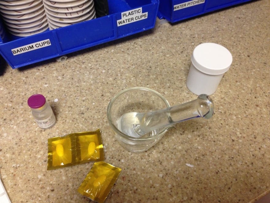
  1. 3 TXA 650 mg tablets
  2. About 2 mL sterile water for injection

Directions

  1. Crush tablets with mortar and pestle and triturate into a fine powder
    • A pill crusher works almost as well if a mortar and pestle are not available
    • The paste can then be prepared in a plastic pill cup
  2. Add sterile water in small aliquots (~0.5 mL) and mix until a thick paste is formed
  3. Apply paste to desired site for 20-30 minutes and remove

tranexamic acid paste

TXA Paste 2

Additional Considerations

  • Use immediately upon compounding. Although oral mouthwashes containing tablets mixed in sterile water are reportedly stable for up to 5 days, stability data for the paste is not available [9].
  • Time to cessation of bleeding after topical TXA has been similar in our experience whether prepared from an IV or PO product.
  • Epistaxis:
    • The paste may not be the best option for epistaxis as it may be difficult to remove all of the paste once applied in the nare(s).
    • Case reports exist for cessation of epistaxis in anticoagulated patients treated with topical IV TXA; however, it is unclear if a TXA paste will have similar results [8].
  • Our institution created an electronic order entry for TXA paste to promote consistency and standardize the process of ordering as well as preparation downstairs in the main pharmacy.
  • There is no published data regarding a topical paste recipe, therefore it is unclear if a smaller dose (e.g. 1-2 tablets) would be as effective as 3 tablets
  • In countries where TXA is supplied as 500 mg tablets, it is reasonable to consider the use of four 500 mg tablets (2000 mg instead of 1950 mg). However, slightly more sterile water may be required to achieve the same consistency as with three 650 mg tablets.

Application to Clinical Practice

  • For patients with minor bleeding not responding to direct pressure, consider topical TXA prior to the use of topical thrombin or more invasive therapies such as injectable lidocaine-epinephrine
  • Locations of minor bleeding in which we have had success include:
    • Post-dental extractions
    • Scalp lacerations
    • Extremity lacerations


References

  1. Zahed R, Moharamzadeh P, Alizadeharasi S, Ghasemi A, Saeedi M. A new and rapid method for epistaxis treatment using injectable form of tranexamic acid topically: a randomized controlled trial. Am J Emerg Med. 2013; 31(9): 1389-92. PMID: 23911102 
  2. Noble S, Chitnis J. Case report: use of topical tranexamic acid to stop localised bleeding. Emerg Med J. 2013; 30(6): 509-10. PMID: 22833592 
  3. Patatanian E, Fugate SE. Hemostatic mouthwashes in anticoagulated patients undergoing dental extraction. Ann Pharmacother. 2006; 40(12): 2205-10. PMID: 17090725 
  4. Waye C. Topical tranexamic acid for epistaxis or oral bleeds. REBEL EM July 2014. http://rebelem.com/topical-tranexamic-acid-epistaxis-oral-bleeds/  
  5. Milne K, Hanel E. SGEM #53: Sunday, Bloody Sunday (Epistaxis and Tranexamic Acid). November 2013. http://thesgem.com/2013/11/sgem53-sunday-bloody-sunday-epistaxis-and-tranexamic-acid/ 
  6. Ambados F. Letter to the editor. Preparing tranexamic acid 4.8% mouthwash. Australian Prescriber. 2003;26:75-77 
  7. Coetzee MJ The use of topical crushed tranexamic acid tablets to control bleeding after dental surgery and from skin ulcers in haemophilia. Haemophilia. 2007; 13(4): 443-4. PMID: 17610565 
  8. Utkewicz MD, Brunetti L, Awad NI. Epistaxis complicated by rivaroxaban managed with topical tranexamic acid. Am J Emerg Med. 2015; 33(9): 1329.e5-7. PMID: 25895714 
  9. Lam MS Extemporaneous compounding of oral liquid dosage formulations and alternative drug delivery methods for anticancer drugs. Pharmacotherapy. 2011; 31(2): 164-92. PMID: 21275495

Author information

Scott Dietrich, PharmD

Scott Dietrich, PharmD

ED Clinical Pharmacist
St. Joseph's Hospital
Tampa, FL

The post Trick of the Trade: Topical Tranexamic Acid Paste for Hemostasis appeared first on ALiEM.


ALiEM Chief Resident Incubator Must Read EM Journal Articles – 2016 Edition

$
0
0

must read em journal articlesIn November 2013, the blog post 52 Articles in 52 weeks: Landmark EM Articles for EM interns was published on the ALiEM site. Over the subsequent years, many ground-breaking and practice-changing articles have been published. As part of a multi-institutional initiative launched by the ALiEM Chief Resident Incubator (“The Crincubator”), 9 chief residents from across the country pooled together lists of journal articles thought to be most important for the broad spectrum of EM learners. Additional input was obtained from FOAM leaders across the country including Dr. Ryan Radecki and Dr. Jeffrey Kline. These lists can be used by individuals for further learning or by residency programs for journal clubs.

Journal Article Selection

These selected journal articles were not restricted to those solely published in the last 2 years, but included groundbreaking articles regardless of their publication date. Lists were then created based on their relevance for junior residents and senior residents/attending physicians. Another list was created which includes controversial hot topics. Each list contains 12 sections to provide a monthly reading list for the individual EM practitioner or longitudinal curriculum for residency programs (e.g. monthly journal clubs). Although these lists are by no means comprehensive, they do cover a large breadth of topics relevant to all EM practitioners.

Special mention contributors: Dr. Casey Hand and Dr. Joe Kennedy

Junior Residents

This compilation of articles was put together to help medical students and junior residents to learn the reasons behind why we practice the way we do in emergency medicine.

Article  Clinical Question
1 Six AJ, Backus BE, Kelder JC. Chest pain in the emergency room: value of the HEART score. Neth Heart J. 2008; 16(6): 191-6. PMID: 18665203 Can the HEART score reliably predict outcomes in chest pain patients?
2 CRASH-2 trial collaborators, Shakur H, Roberts I, et al. Effects of tranexamic acid on death, vascular occlusive events, and blood transfusion in trauma patients with significant haemorrhage (CRASH-2): a randomised, placebo-controlled trial. Lancet. 2010; 376(9734): 23-32. PMID: 20554319 Should tranexamic acid be considered in the management of trauma patients with significant hemorrhage?
3 Kuppermann N, Holmes JF, Dayan PS, et al. Identification of children at very low risk of clinically-important brain injuries after head trauma: a prospective cohort study. Lancet. 2009; 374(9696): 1160-70. PMID: 19758692 When is a head CT indicated in children with blunt head trauma? [PV card]
4 Hoffman JR, Mower WR, Wolfson AB, Todd KH, Zucker MI. Validity of a set of clinical criteria to rule out injury to the cervical spine in patients with blunt trauma. National Emergency X-Radiography Utilization Study Group. N Engl J Med. 2000; 343(2): 94-9. PMID: 10891516 What criteria can be used to rule out cervical spine injury without imaging? How were these criteria defined in the study?
5 Stiell IG, Clement CM, McKnight RD, et al. The Canadian C-spine rule versus the NEXUS low-risk criteria in patients with trauma. N Engl J Med. 2003; 349(26): 2510-8. PMID: 14695411 How does the Canadian C-Spine rule compare with the NEXUS criteria for ruling out cervical spine injury?
6 Stiell IG, Clement CM, Rowe BH, et al. Comparison of the Canadian CT Head Rule and the New Orleans Criteria in patients with minor head injury. JAMA. 2005; 294(12): 1511-8. PMID: 16189364 What are the criteria for clinically ruling out head injury in patients with blunt head trauma? How does the Canadian CT Head Rule compare with the New Orleans Criteria?
7

Stiell IG, Greenberg GH, McKnight RD, et al. Decision rules for the use of radiography in acute ankle injuries. Refinement and prospective validation. JAMA. 1993; 269(9): 1127-32. PMID: 8433468


Stiell IG, Greenberg GH, Wells GA, et al. Prospective validation of a decision rule for the use of radiography in acute knee injuries. JAMA. 1996; 275(8): 611-5. PMID: 8594242

What criteria can be used to rule out significant foot, ankle, and knee injuries without imaging? [PV card]
8 Sharifi M, Bay C, Skrocki L, Rahimi F, Mehdipour M, “MOPETT” Investigators. Moderate pulmonary embolism treated with thrombolysis (from the “MOPETT” Trial). Am J Cardiol. 2013; 111(2): 273-7. PMID: 23102885 Should “safe dose” tPA be given to patients with submassive pulmonary embolism? Is it safe? How efficacious is it?
9

ProCESS Investigators, Yealy DM, Kellum JA, et al. A randomized trial of protocol-based care for early septic shock. N Engl J Med. 2014; 370(18): 1683-93. PMID: 24635773


ARISE Investigators, ANZICS Clinical Trials Group, Peake SL, et al. Goal-directed resuscitation for patients with early septic shock. N Engl J Med. 2014; 371(16): 1496-506. PMID: 25272316


Mouncey PR, Osborn TM, Power GS, et al. Trial of early, goal-directed resuscitation for septic shock. N Engl J Med. 2015; 372(14): 1301-11. PMID: 25776532

In the current management of sepsis, does early goal-directed therapy improve outcomes compared with usual care?
10 Holst LB, Haase N, Wetterslev J, et al. Lower versus higher hemoglobin threshold for transfusion in septic shock. N Engl J Med. 2014; 371(15): 1381-91. PMID: 25270275 What hemoglobin threshold should be used for transfusing septic patients?
11 Weingart SD, Levitan RM. Preoxygenation and prevention of desaturation during emergency airway management. Ann Emerg Med. 2012; 59(3): 165-75.e1. PMID: 22050948 What techniques can be utilized to improve preoxygenation and prevent desaturation in the peri-intubation setting?
12 Pickard R, Starr K, MacLennan G, et al. Medical expulsive therapy in adults with ureteric colic: a multicentre, randomised, placebo-controlled trial. Lancet. 2015; 386(9991): 341-9. PMID: 25998582 Are tamsulosin or nifedipine more effective than placebo for improving ureteral stone passage?


Senior Residents and Attendings

This list was designed with slightly more advanced topics that are best suited for those further along in their training.

Article  Clinical Question
1 Wyse DG, Waldo AL, DiMarco JP, et al. A comparison of rate control and rhythm control in patients with atrial fibrillation. N Engl J Med. 2002; 347(23): 1825-33. PMID: 12466506 Which will help a patient with atrial fibrillation more? Rate or rhythm control?
2 Anderson CS, Heeley E, Huang Y, et al. Rapid blood-pressure lowering in patients with acute intracerebral hemorrhage. N Engl J Med. 2013; 368(25): 2355-65. PMID: 23713578 Does standard, or rapid, lowering of blood pressure in acute intracranial hemorrhage improve outcomes?
3 Delaney KA, Vassallo SU, Larkin GL, Goldfrank LR. Rewarming rates in urban patients with hypothermia: prediction of underlying infection. Acad Emerg Med. 2006; 13(9): 913-21. PMID: 16946289 What is the underlying cause of a patient’s hypothermia? Could s/he be septic?
4 Newman-Toker DE, Kerber KA, Hsieh YH, et al. HINTS outperforms ABCD2 to screen for stroke in acute continuous vertigo and dizziness. Acad Emerg Med. 2013; 20(10): 986-96. PMID: 24127701 Should I use the ABCD2 score or the HINTS exam to determine the likelihood of stroke in a dizzy patient?
5 Leuppi JD, Schuetz P, Bingisser R, et al. Short-term vs conventional glucocorticoid therapy in acute exacerbations of chronic obstructive pulmonary disease: the REDUCE randomized clinical trial. JAMA. 2013; 309(21): 2223-31. PMID: 23695200 Should I send my COPD patient home on a 14 day or 4 day course of oral steroids?
6 Goyal M, Demchuk AM, Menon BK, et al. Randomized assessment of rapid endovascular treatment of ischemic stroke. N Engl J Med. 2015; 372(11): 1019-30. PMID: 25671798 Do I need to call the neurointerventionalist to retrieve this clot in my patient with an ischemic stroke?
7 Berkhemer OA, Fransen PS, Beumer D, et al. A randomized trial of intraarterial treatment for acute ischemic stroke. N Engl J Med. 2015; 372(1): 11-20. PMID: 25517348 Does intraarterial treatment of ischemic stroke improve outcomes?
8 Villanueva C, Colomo A, Bosch A, et al. Transfusion strategies for acute upper gastrointestinal bleeding. N Engl J Med. 2013; 368(1): 11-21. PMID: 23281973 Should I transfuse my patient with a hemoglobin of 7 g/dL (restrictive strategy) or 9 g/dL (liberal strategy) in the setting of a gastrointestinal bleed? 
9 Easter JS, Haukoos JS, Claud J, et al. Traumatic intracranial injury in intoxicated patients with minor head trauma. Acad Emerg Med. 2013; 20(8): 753-60. PMID: 24033617 Do my traditional head injury decision rules apply to the intoxicated patient population?
10 Salminen P, Paajanen H, Rautio T, et al. Antibiotic Therapy vs Appendectomy for Treatment of Uncomplicated Acute Appendicitis: The APPAC Randomized Clinical Trial. JAMA. 2015; 313(23): 2340-8. PMID: 26080338 Should your patient with appendicitis just receive a trial of antibiotics instead of surgery as a first line approach?
11 Mal S, McLeod S, Iansavichene A, Dukelow A, Lewell M. Effect of out-of-hospital noninvasive positive-pressure support ventilation in adult patients with severe respiratory distress: a systematic review and meta-analysis. Ann Emerg Med. 2014; 63(5): 600-607.e1. PMID: 24342819 Does use of NIPPV (noninvasive positive pressure ventilation) in the pre-hospital setting improve outcomes in acute respiratory distress patients?
12 Silbergleit R, Durkalski V, Lowenstein D, et al. Intramuscular versus intravenous therapy for prehospital status epilepticus. N Engl J Med. 2012; 366(7): 591-600. PMID: 22335736 Does route of benzodiazepine administration change time to seizure control in pre-hospital status epilepticus?


Controversial Topics

The collection was put together with controversial, novel, or potentially practice changing topics to help us think about new things on the horizon and stimulate discussion amongst colleagues.  

Article  Clinical Question
1 Inaba K, Lustenberger T, Recinos G, et al. Does size matter? A prospective analysis of 28-32 versus 36-40 French chest tube size in trauma. J Trauma Acute Care Surg. 2012; 72(2): 422-7. PMID: 22327984 Does chest tube size in traumatic injury change outcomes and patient satisfaction?
2 Isacson D, Thorisson A, Andreasson K, Nikberg M, Smedh K, Chabok A. Outpatient, non-antibiotic management in acute uncomplicated diverticulitis: a prospective study. Int J Colorectal Dis. 2015; 30(9): 1229-34. PMID: 25989930 Should this uncomplicated acute diverticulitis patient be given oral antibiotics?
3 Marik PE, Flemmer M, Harrison W. The risk of catheter-related bloodstream infection with femoral venous catheters as compared to subclavian and internal jugular venous catheters: a systematic review of the literature and meta-analysis. Crit Care Med. 2012; 40(8): 2479-85. PMID: 22809915 Is my patient at higher risk of infection if I place a femoral central venous catheter over an internal jugular or subclavian catheter?
4 Nielsen N, Wetterslev J, Cronberg T, et al. Targeted temperature management at 33°C versus 36°C after cardiac arrest. N Engl J Med. 2013; 369(23): 2197-206. PMID: 24237006 My post-cardiac arrest patient needs to be cooled, do I cool to 33°C, or is 36°C sufficient?  Will this change my patient’s outcome?
5 Stub D, Smith K, Bernard S, et al. Air Versus Oxygen in ST-Segment-Elevation Myocardial Infarction. Circulation. 2015; 131(24): 2143-50. PMID: 26002889 In medical school I learned about MONA therapy for all of my STEMI patients.  Do they still need oxygen even if they are not hypoxic? Does this improve my patient’s chance of a good outcome?
6 McCormack RF, Hutson A. Can computed tomography angiography of the brain replace lumbar puncture in the evaluation of acute-onset headache after a negative noncontrast cranial computed tomography scan? Acad Emerg Med. 2010; 17(4): 444-51. PMID: 20370785 I am concerned my patient has a subarachnoid hemorrhage as the cause of his headache, do I need to perform a lumbar puncture to prove/disprove this?
7 Semler MW, Janz DR, Lentz RJ, et al. Randomized Trial of Apneic Oxygenation during Endotracheal Intubation of the Critically Ill. Am J Respir Crit Care Med. 2016; 193(3): 273-80. PMID: 26426458 Will apneic oxygenation during intubation prevent my patient from desaturating early in the procedure?
8 Grunau BE, Wiens MO, Rowe BH, et al. Emergency Department Corticosteroid Use for Allergy or Anaphylaxis Is Not Associated With Decreased Relapses. Ann Emerg Med. 2015; 66(4): 381-9. PMID: 25820033 Will the addition of corticosteroids in my anaphylactic patient help to prevent future allergy relapses?
9 Righini M, Van Es J, Den Exter PL, et al. Age-adjusted D-dimer cutoff levels to rule out pulmonary embolism: the ADJUST-PE study. JAMA. 2014; 311(11): 1117-24. PMID: 24643601 Can I adjust my D-dimer for my patient’s age to aid in ruling-out pulmonary embolism? What about in pregnancy?
10

Moore CL, Bomann S, Daniels B, et al. Derivation and validation of a clinical prediction rule for uncomplicated ureteral stone–the STONE score: retrospective and prospective observational cohort studies. BMJ. 2014; 348: g2191. PMID: 24671981


Smith-Bindman R, Aubin C, Bailitz J, et al. Ultrasonography versus computed tomography for suspected nephrolithiasis. N Engl J Med. 2014; 371(12): 1100-10. PMID: 25229916

Can I avoid unnecessarily irradiating my renal colic patient and diagnose nephrolithiasis with ultrasound? Is there a decision rule I can use to help determine the likelihood that my patient has a kidney stone?
11 Beam DM, Kahler ZP, Kline JA. Immediate Discharge and Home Treatment With Rivaroxaban of Low-risk Venous Thromboembolism Diagnosed in Two U.S. Emergency Departments: A One-year Preplanned Analysis. Acad Emerg Med. 2015; 22(7): 788-95. PMID: 26113241 Can I send my newly diagnosed venous thromboembolism patient home with oral anticoagulation and outpatient follow up?
12 Schwarz ES, Cohn BG. Is dexamethasone as effective as prednisone or prednisolone in the management of pediatric asthma exacerbations? Ann Emerg Med. 2015; 65(1): 81-2. PMID: 24954577 Getting kids to take medication can be so difficult sometimes. Could a single home dose of dexamethasone be sufficient to treat a pediatric asthma exacerbation?


Image (c) Can Stock Photo

Author information

Allison Trop, MD

Allison Trop, MD

Emergency Medicine Chief Resident
John Peter Smith Health Network

The post ALiEM Chief Resident Incubator Must Read EM Journal Articles – 2016 Edition appeared first on ALiEM.

60 Second Soapbox: Bafuma (IV Antibiotics), Favot (Nod and Smile), & Augustine (Medical Travel)

$
0
0

60-second soapboxGet ready for another round of 60-Second Soapbox! Each episode, one lucky individual gets exactly 1 minute to present their rant-of-choice to the world. Any topic is on the table – clinical, academic, economic, or whatever else may interest an EM-centric audience. We carefully remix your audio to add an extra splash of drama and excitement. Even more exciting, participants get to challenge 3 of their peers to stand on a soapbox of their own!

bafuma

Patrick Bafuma, PA-C
@EMinFocus
Lead Physician Assistant, Columbia Memorial Hospital

#FutureSoapboxChallenge

References

  1. Chan R. Oral versus intravenous antibiotics for community acquired lower respiratory tract infection in a general hospital: open, randomised controlled trial. BMJ. 1995. PMID: 7787537
  2. Haran JP. Factors influencing the development of antibiotic associated diarrhea in ED patients discharged home: risk of administering IV antibiotics. Am J Emerg Med. 2014. PMID : 25149599
  3. Lorgelly PK. Oral versus i.v. antibiotics for community-acquired pneumonia in children: a cost-minimisation analysis. Eur Respir J. 2010. PMID: 19717479
favot_circle

Dr. Mark Favot
@ten8suited
Emergency Ultrasound Fellowship Director
Wayne State University/Sinai-Grace Hospital

#FutureSoapboxChallenge

augustine

Dr. Tamara Augustine
Chief Resident, Wayne State University/Sinai-Grace Hospital
Matriculating Global Health Fellow, University of California – San Francisco

#FutureSoapboxChallenge

  • Dr. Katrina Godderz
  • Dr. Ciara Barclay-Buchanan (@cbb_md)
  • Dr. Karyn Kohler

Author information

Sam Shaikh, DO

Sam Shaikh, DO

Editor, 60-Second Soapbox series
Emergency Medicine Chief Resident
Sinai-Grace Hospital/ Detroit Medical Center
2014-15 ALiEM-CORD Social Media and Digital Scholarship Fellow

The post 60 Second Soapbox: Bafuma (IV Antibiotics), Favot (Nod and Smile), & Augustine (Medical Travel) appeared first on ALiEM.

Fentanyl: Adding Fuel to the Fire in the North American Opioid Epidemic

$
0
0

FentanylDrug poisoning is now the leading cause of injury death in the United States [1], with opioids accounting for up to 40% of these deaths. In the U.S., prescription opioid death rates have more than quadrupled since 1999, and death rates exceed those due to motor vehicle crashes [2]. Similar trends in opioid exposure and death rates in Canada suggest that it is not far behind. Prescriptions for opioid analgesics paralleled a rise in opioid abuse and fatalities between 2002 and 2010, leveling off between 2011 and 2013 [3], only to rise again in 2014 [1]. Among the more frequently misused opioids nationwide are oxycodone and hydrocodone (the most widely prescribed drug in the U.S.) in their various formulations, and methadone, but a “rising star” in the epidemic in many regions is fentanyl.

In March 2015, the U.S. Drug Enforcement Administration (DEA) issued nationwide alerts that identified fentanyl as a significant threat to public health and safety [10]. The National Forensic Laboratory Information System report a more than 8-fold increase in the total number of fentanyl seizures between 2012 and 2014 [11]. Additionally, in late 2013 and 2014, the DEA National Heroin Threat Assessment Summary noted spikes in overdose deaths related to fentanyl and its analog, acetyl-fentanyl, throughout the U.S. [12]

What is fentanyl? Is it any worse than other opioids?

Fentanyl is a commonly used medication in Emergency Departments (EDs) and inpatient units throughout Canada and the U.S. It is effective for pain control when used appropriately and is about 100 times more potent than morphine and 30-50 times more potent than heroin. Fentanyl is a schedule I drug by the Canadian Controlled Drugs and Substances Act and schedule II by the U.S. Controlled Substances Act. This scheduling reinforces that although fentanyl has currently accepted medical uses, it carries a high potential for abuse and may lead to severe psychological or physical dependence.

Pharmaceutical fentanyl is available in injectable solutions (intravenous and intramuscular), transdermal patches, sublingual sprays, transmucosal lozenges, transbuccal tablets, and intranasal sprays. Access to pharmaceutical fentanyl for the purpose of abuse occurs primarily through diversion or theft of prescribed fentanyl in transdermal formulation.

However, since much of the fentanyl now available for abuse is illicitly manufactured, it is referred to as non-pharmaceutical fentanyl (NPF). Although large-scale, epidemic fentanyl and fentanyl analog (i.e. acetyl-fentanyl, α-methylfentanyl) poisoning is relatively new, small outbreaks have occurred numerous times in the U.S. over the past 25 years [4][5][6][7][8][9]. Testing of drug samples is able to differentiate between pharmaceutical grade fentanyl and NPF, and the majority of the >700 fentanyl-related overdose deaths during this time were attributed to NPF [11].

Fentanyl powder is highly concentrated and requires dilution before it is combined with other street drugs, including heroin, or pressed into pills and sold on the streets. Common routes of exposure include intravenous, oral, and insufflation. Despite the poor oral bioavailability of fentanyl, it is easy to administer sufficient amounts of drug to allow absorption of sufficient amounts to cause euphoria, as well as respiratory depression and death. The amount of drug, and even the specific content of the product being used (e.g., could it be a fentanyl analog) is essentially unknowable by the consumer. Combined with the extreme potency of fentanyl and its derivatives (some 10,000 times that of morphine), the risk of overdose is extremely high.

How and where is NPF being produced? How is NPF used?

NPF and fentanyl analogs are produced in clandestine laboratories through relatively simple chemical processes that can be found online. Most commonly, NPF is produced via the Siegfried method that uses N-phenethyl-piperidone as a precursor. In 2006, in response to rising numbers of NPF-related deaths, the CDC implemented an ad-hoc case finding and surveillance system that identified 1,013 NPF-related deaths between April 4, 2005 and March 28, 2007. The production of NPF was related to clandestine production in a laboratory in Toluca, Mexico, as well as domestic production within the U.S. [13] As a result, in 2007 the DEA began regulating access to N-phenethyl-piperidone [14].

Currently, clandestinely-produced fentanyl is primarily sourced from Mexico; fentanyl analogs and precursor chemicals are obtained from distributors in other countries including China, Germany, and Japan [10]. Pharmaceutical fentanyl is also diverted for abuse, but at much lower levels. There have also been several clandestine laboratories identified in Canada by the Royal Canadian Mounted Police (RCMP), however, much of the NPF is produced in other countries, predominantly China, then imported to Canada.

Of note, on October 1, 2015, the Chinese Ministry of Public Security (MPS) Narcotics Control Bureau announced the regulation of the sale and distribution of 116 chemical compounds used in the production of synthetic drugs, including acetyl-fentanyl. Chinese officials declared these compounds were found to have no known legitimate use and therefore will be controlled administratively by the MPS.

What is the epidemiology of fentanyl use in North America?

Over the past several years, many provinces across Canada, including Alberta, have seen a significant increase in exposures and deaths related to fentanyl and fentanyl analog exposure. Illicit NPF was first identified in Montreal, Quebec in May 2013, and the Canadian Community Epidemiology Network on Drug Use (CCENDU) correctly predicted that the availability of NPF was likely to spread throughout Canada to other regions. CCENDU report 1,019 drug poisoning deaths between 2009 and 2014 in whom post-mortem toxicology analysis identified fentanyl.

Since June 2013, the Canadian Centre on Substance Abuse (CCSA) has reported the availability of NPF in pill and powder form across Canada that is being sold on the street as “green monsters”, “green beans”, “green jellies”, or “street oxy”. It is often sold to users who believe they are receiving oxycodone, heroin, or other drugs of abuse including stimulant drugs such as MDMA (“ecstasy”) and cocaine [15]. In fact, many pills being sold as counterfeit oxycodone  are made to resemble Oxycontin® or Roxicodone, with identical markings and colors, prompting stricter regulation over the purchase and use of pill-forming machines. Health Canada’s Drug Analysis Service has identified fentanyl in 89% of samples of seized counterfeit oxycodone tablets, and overall seizures of fentanyl, including both diverted prescription grade and NPF, increased over 30 times between 2009 and 2014 [16].

NPF drug seizures and deaths have had a similar progress across the U.S., particularly the eastern half [11]. Rhode Island experienced 12 deaths in 2013 from acetylfentanyl [8].

In the St. Louis area, fentanyl alone was the primary death factor in 44% of fentanyl-related overdose cases while the remaining 56% involved fentanyl along with other substances such as alcohol, pharmaceuticals, cocaine, or heroin.

In Alberta specifically, with a population of roughly 4 million, there were 162 fentanyl-detected deaths (including all deaths for which post-mortem toxicological testing detected the presence of fentanyl) and 61 fentanyl-implicated deaths (where fentanyl was identified as the direct cause or a contributing cause of death) between 2011 and 2014 [16]. The scope of the epidemic is so great that Alberta’s health authority has formed an ad hoc emergency command center, previously reserved for infectious disease outbreaks such as measles or H1N1, to coordinate efforts against the rising public health threat.

In July 2015, the DEA New Jersey Tactical Diversion Squad identified an illicit organization that was distributing counterfeit Roxicodone pills that were in fact 40% acetyl-fentanyl. Subsequent testing of similar Roxicodone pills from the same organization in December 2015 revealed that the pills now contained 60% pharmaceutical grade fentanyl-citrate. Not surprisingly, many chronic opioid abusers who present with unintentional fentanyl overdoses believed they were purchasing oxycodone or another drug of abuse, and the significantly different potency and pharmacokinetics of fentanyl contributed to opioid toxicity. However, not all fentanyl use occurs in chronic opioid users, and many overdoses occur in opioid-naïve users under the age of 40 [17].

News reports of fentanyl abuse in the United States
Fake Fentanyl Pills Led To Hundreds of Overdose Deaths in Cuyahoga County (Cleveland)
Fentanyl: drug 50 times more potent than heroin ravages New Hampshire.
Dangerous cancer drug on the rise in Louisville
Fentanyl overdoses in New Orleans reflect national trend
Officials lack clear link to fentanyl in heroin deaths
La Crosse (Wisconsin) health officials warn about dangerous Fentanyl laced heroin
Fentanyl: Little known drug causing deaths to skyrocket in Palm Beach County (Florida)


Map canstockphoto3179047

What are the challenges in tracking the fentanyl epidemic?

Several limitations regarding exposure and death reporting make estimating the overall burden on society difficult. First, many patients with fentanyl exposures who present to the ED are unidentified as such (i.e., fentanyl cannot be detected by a hospital laboratory; it is very hard to detect the analogues even in a reference laboratory) and remain unreported to the regional poison control centers and health departments, leading to underrepresentation of the incidence of exposures. Without accurate history from family or friends, these cases may not be appreciated as fentanyl related. The medical examiner/coroners suffer the same limitations.

The commonly accepted response to naloxone as diagnostic of an opioid overdose may not occur in patients with late presentations to healthcare following fentanyl exposure since they have already developed sequelae of prolonged hypoxic injury (this is true for all opioids). However, it is especially concerning with fentanyl, given its high potency and the ease in which a profound overdose can occur. Also, drug poisoning death reporting and certification varies across regions throughout North America. Furthermore, although in the U.S. the National Center for Health Statistics tracks fentanyl-related deaths through the use of death certificates, a national surveillance system is currently lacking in Canada. The Statistics Canada Vital Statistics Death Database offers a potential source for estimating opioid related deaths, however its utility relies on accurate coding of patient presentations using International Statistical Classification of Diseases, 10th revision (ICD-10), which, as noted, is limited by the ability to make the clinical diagnosis [18].

What is the treatment for patients with fentanyl overdose?

Naloxone is a competitive opioid antagonist, capable of reversing the effects of fentanyl and other opioid agonists. Patients in the ED with respiratory depression from suspected opioid toxicity, who are not intubated, are candidates for the administration of naloxone. The starting IV dose is 0.04 mg, and this is titrated to effect [19]. Most patients with opioid-induced respiratory depression will respond to small doses of naloxone [20][21]; however, anecdotally, some report that patients with fentanyl poisoning have required very high doses of fentanyl (up to 12 mg) to reverse opioid effects [9]. Because there is no pharmacologic or clinical reason to explain this requirement, it may be due to the use of a fentanyl analog (with high binding affinity for the mu receptor), another concomitant sedative, hypoxia, or a nonspecific effect.

Naloxone administration is predominantly done by trained health care providers, including ED physicians and EMS personnel. Given the rapid and potent effects of fentanyl, significant hypoxia and end organ effects may occur by the time EMS is activated; the ability to administer an opioid antagonist early after overdose offers significant potential benefits to morbidity and mortality.

Take-home naloxone programs

Take-home naloxone programs have been working to deliver naloxone training to high-risk inpatients, ED patients, and outpatients seen in clinic, as well as to friends and family who may be present during an opioid overdose [14]. These programs first appeared in the U.S. in 1996 and have continued to grow and expand, with 9 states passing laws to expand access to naloxone in early 2015, taking the total number of jurisdictions with naloxone access laws to 37 as of July 2015 [22][23]. Programs are sprouting up across Canada as well. A survey from the Harm Reduction Coalition assessed organizations in the US distributing naloxone kits between 1996 and June 2014 and found that 152,283 kits were distributed with reports of 26,463 administrations [22].

In addition to these community-based distribution programs for take home naloxone, New York City has now made naloxone available for purchase over the counter, further increasing availability of the potentially life saving antidote. Others are following this trend, and Health Canada has made similar proposals to allow access to naloxone without a prescription.

What does the future hold?

The fentanyl epidemic across Canada and the U.S. is a significant threat to public health. The development of this epidemic has paralleled that of heroin, and it may be that a similar demographic of former prescription opioid users are at greatest risk [24][25]. The meteoric rise in use and consequence shows no current signs of slowing and reinforces the need for improved methods for identification and reporting of fentanyl exposures and deaths. In the background of the fentanyl epidemic are a new class of ultrapotent opioids called the research chemical (RC) opioids. Drugs such as W-18 have potencies that dwarf those of the fentanyls and are very difficult to detect analytically. We hope that through public education, intelligence, and law enforcement efforts, the expanded use of take-home naloxone, and support of addiction services will help to curb the use and deaths from of fentanyl abuse.

References

 
  1. Warner M, Chen LH, Makuc DM, Anderson RN, et al. Drug poisoning deaths in the United States, 1980-2008. NCHS Data Brief 2011 (81):1-8. 
  2. Chen LH, Hedegaard H, Warner M. Drug-poisoning Deaths Involving Opioid Analgesics: United States, 1999-2011. NCHS Data Brief 2014 (166):1-8. 
  3. Dart RC, Severtson SG, Bucher-Bartelson B. Trends in opioid analgesic abuse and mortality in the United States. N Engl J Med. 2015; 372(16): 1573-4. PMID: 25875268 
  4. Martin M, Hecker J, Clark R, et al. China White epidemic: an eastern United States emergency department experience. Ann Emerg Med. 1991; 20(2): 158-64. PMID: 1996799 
  5. Hibbs J, Perper J, Winek CL. An outbreak of designer drug–related deaths in Pennsylvania. JAMA. 1991; 265(8): 1011-3. PMID: 1867667
  6. Acetyl fentanyl overdose fatalities–Rhode Island, March-May 2013. MMWR Morb Mortal Wkly Rep 2013;62(34):703-4. 
  7. Algren DA, Monteilh CP, Punja M, et al. Fentanyl-associated fatalities among illicit drug users in Wayne County, Michigan (July 2005-May 2006). J Med Toxicol. 2013; 9(1): 106-15. PMID: 23359211
  8. Mercado-Crespo MC, Sumner SA, Spelke MB, Sugerman DE, et al. Notes from the field: increase in fentanyl-related overdose deaths – Rhode Island, November 2013-March 2014. MMWR Morb Mortal Wkly Rep 2014;63(24):531.  
  9. Schumann H, Erickson T, Thompson TM, Zautcke JL, Denton JS. Fentanyl epidemic in Chicago, Illinois and surrounding Cook County. Clin Toxicol (Phila). 2008; 46(6): 501-6. PMID: 18584361
  10. US Drug Enforcement Administration. DEA Issues Nationwide Alert on Fentanyl as Threat to Health and Public Safety. March 2015. Accessed December 15, 2015. 
  11. CDC Health Alert Network. Increases in Fentanyl Drug Confiscations and Fentanyl-related Overdose Fatalities. October 2015. Accessed December 15, 2015. 
  12. US Drug Enforcement Administration Intelligence Report. National Heroin Threat Assessment Summary (PDF). April 2015. Accessed December 15, 2015. 
  13. US Drug Enforcement Administration. Control of a Chemical Precursor Used in the Illicit Manufacture of Fentanyl as a List I Chemical. April 2007. Accessed January 4, 2015. 
  14. Nonpharmaceutical fentanyl-related deaths–multiple states, April 2005-March 2007. MMWR Morb Mortal Wkly Rep 2008;57(29):793-6. 
  15. CCENDU Drug Alert: Illicit Fentanyl (PDF). 2013. Accessed October 25, 2015. 
  16. CCENDU Bulletin: Deaths Involving Fentanyl in Canada, 2009-2014 (PDF). 2015. Accessed October 25, 2015. 
  17. CCENDU Drug Alert: Fentanyl-related Overdoses (PDF). 2015. Accessed October 25, 2015. 
  18. Gladstone E, Smolina K, Morgan SG, Fernandes KA, Martins D, Gomes T. Sensitivity and specificity of administrative mortality data for identifying prescription opioid-related deaths. CMAJ. 2015. PMID: 26622006 Kim HK, Nelson LS. Reversal of Opioid-Induced Ventilatory Depression Using Low-Dose Naloxone (0.04 mg): a Case Series. J Med Toxicol. 2015. PMID: 26289651 
  19. Takahashi M, Sugiyama K, Hori M, Chiba S, Kusaka K. Naloxone reversal of opioid anesthesia revisited: clinical evaluation and plasma concentration analysis of continuous naloxone infusion after anesthesia with high-dose fentanyl. J Anesth. 2004; 18(1): 1-8. PMID: 14991468
  20. Tigerstedt I Reversal of fentanyl-induced narcotic depression with naloxone following general anaesthesia. Acta Anaesthesiol Scand. 1978; 22(3): 234-40. PMID: 676644
  21. Wheeler E, Jones TS, Gilbert MK, Davidson PJ. Opioid Overdose Prevention Programs Providing Naloxone to Laypersons – United States, 2014. MMWR Morb Mortal Wkly Rep 2015;64(23):631-5. 
  22. Naloxone overdose prevention laws. Prescription Drug Abuse Policy System (July 2015). Accessed December 5, 2015. 
  23. Cicero TJ, Ellis MS, Harney J. Shifting Patterns of Prescription Opioid and Heroin Abuse in the United States. N Engl J Med. 2015; 373(18): 1789-90. PMID: 26510045
  24. Compton WM, Jones CM, Baldwin GT. Relationship between Nonmedical Prescription-Opioid Use and Heroin Use. N Engl J Med. 2016; 374(2): 154-63. PMID: 26760086
  25. Rudd RA, Aleshire N, Zibbell JE, Gladden RM. Increases in Drug and Opioid Overdose Deaths – United States, 2000-2014. MMWR Morbidity and Mortality Weekly Report. 2016;64(50-51):1378–82.

Further Reading:

  • Interactive.fusion.net site. Death by Fentanyl (2016). Accessed February 23, 2016.


Images: NaloxoneMap (c) Can Stock Photo

Author information

Scott Lucyk, MD

Scott Lucyk, MD

Emergency physician, Medical Toxicologist
Department of Emergency Medicine
University of Calgary, Alberta Health Services
Poison and Drug Information Service (PADIS)

The post Fentanyl: Adding Fuel to the Fire in the North American Opioid Epidemic appeared first on ALiEM.

PEM Pearls: Pediatric Concussions

$
0
0

pediatric concussions canstockphoto0691281A 9-year boy was hit in the head during a soccer game and was out for a few seconds. He regained consciousness quickly, but was repetitive for EMS. By the time the patient arrived at the ED, he was back to his normal self. Did this patient sustain a concussion? If so, what discharge instructions, anticipatory guidance, and resources do you have for your patient and his family? Here’s a quick 170-second animated video tutorial to sum up some thing for you.


Animated Video on Pediatric Concussions

Background about Pediatric Concussions

About 1.6-3.8 million sports-related concussions occur yearly and account for 10% of sports-related injuries with many of these children presenting to the ED after the accident [1][2]. Boutis et al. found that ED physicians tend to under-diagnose concussions [3]. Common post-concussive symptoms include any of the following symptoms:

  • Headache
  • Confusion
  • Inattentiveness
  • Photophobia
  • Nausea
  • Tiredness

The Zurich Consensus developed guidelines (Sports Concussion Assessment Tool) to assist providers in determining if a child has had a concussion [4]

Patient and Family Education

Worried parents may be concerned about chronic brain injury and will want to know what are the next steps to protect their child. The current recommendations encourage both cognitive and physical rest, followed by graduated return to activity. However, it is unclear what is the optimal balance between rest and play. The Zurich Consensus recommends that children should not return to play on the same day [4]. A randomized controlled study by Thomas et al. found that patients who had strict rest for 5 days had slower resolution of their symptoms compared to those who were allowed to have a more liberalized recovery plan. Furthermore, while both groups had comparable return times to physical activity, those in the strict rest group had longer emotional symptoms, such as depression, compared to those that were in the control group [5].

While most ED physicians are well-versed in the initial evaluation of pediatric head injury, we may be less familiar with resources and recommendations for care after concussion. Several models have been created for individualized treatment plans that take a multidisciplinary approach. The CDC has a website, called Heads Up, which provides resources for parents, coaches and providers. The ED assessment tool, Acute Concussion Evaluation, can help ED doctors objectively evaluate the patient and provide an initial medical assessment that can be used comparatively at the follow-up pediatrician visit. There is also a useful printout for discharge instructions to provide to parents. The California Interscholastic Federation (CIF) provides informational sheets (available in Spanish) and a letter-to-school form.

The Center for Concussion in Colorado created an integrative model, REAP (Remove-Reduce/Educate/Adjust-Accommodate/Pace). This well-coordinated program includes the initial ED evaluation and educational handouts for parents, as well as linked communication between the follow-up clinic and school to notify them that a post-concussive student has been evaluated [1].

Most patients with concussive symptoms will improve within 7-10 days, while a majority will be asymptomatic after 3 weeks. About 10% will continue to be symptomatic after 3 months [4][6]. Follow up should be arranged within a few days for re-evaluation. Some patients may have delayed onset or worsening of symptoms, therefore, it is important to address parental expectations and review strict return precautions (eg. the worst headache in their life, focal neurologic deficits, seizures, and repetitive vomiting).

Returning to School

Children do not need to have complete resolution of symptoms prior to returning back to school, but it is important to excuse children from over-doing homework and/or to notify teachers that the child will need to take frequent breaks. Some non-contact light exercise can also help in persistent symptomatic cases, though this should be discussed with the pediatrician. The Zurich Consensus also created a Graduated Return-to-Play Protocol that can be used to as a guideline (Table 1). If at any point, the patient develops symptoms, then it is recommended to back off and rest. Most states have laws that mandate medical clearance to return back to sports.

Table 1. Graduated return to play protocol [4]

Rehabilitation Stage Functional exercise at each stage of rehabilitation
1. No activity Symptom limited physical and cognitive rest
2. Light aerobic exercise Walking, swimming, stationary cycling, 70% of max heart rate
3. Sport-specific exercise Can do drills but no head impact activity (e.g. running drills in soccer)
4. Non-contact training drills More complex training drills (e.g. passing drills in football)
5. Full-contact practice Following medical clearance, can participate in normal training
6. Return to play Normal game play

Bottom Line

While the pathophysiology of concussions are not well-understood, the detrimental effects on cognitive and physical recovery has been frequently noted if a patient sustains another head injury too soon, AKA “The 2nd Hit”! ED providers are frequently the first physicians to evaluate the patient and provide guidance to the family. It is safe for children to return to school and non-contact play in a graduated process. There are many programs or resources available on post-concussive care. Link patients to appropriate follow-up care after a concussion and remind them that they will need medical clearance to return to play.

References

  1. Kirelik SB, Acute Concussion Management with Remove-Reduce/Educate/Adjust-Accommodate/Pace (REAP). J Emerg Med. 2015 Nov 14, PMID: 26589561.
  2. Meehan WP, Bachur RG. The recommendation for rest following acute concussion. Pediatrics. 2015 Feb;135(2):362-3 PMID: 25560439. 
  3. Boutis K, The diagnosis of concussion in a pediatric emergency department J Pediatr. 2015 May;166(5):1214-1220 PMID: 25919731. 
  4. McCrory P, Consensus statement on Concussion in Sports- The 4th International Conference on Concussion in Sport held in Zurich, November 2012,  Phys Ther Sport. 2013 May;14(2):e1-e13. PMID: 23664041. 
  5. Thomas DG, Benefits of strict rest after acute concussion: a randomized controlled trial., Pediatrics. 2015 Feb;135(2):213-23 PMID: 25560444. 
  6. Interview with Dr. Carlin Senter, UCSF Concussion Clinic, December 7th, 2015 


Image (c) Can Stock Photo

Author information

Delphine Huang, MD

Delphine Huang, MD

Emergency Medicine resident

UCSF-San Francisco General Hospital Residency Program

The post PEM Pearls: Pediatric Concussions appeared first on ALiEM.

AIR Series: Neurology Module 1 – Bleeds and Strokes

$
0
0

ALiEM-AIR-Badge only 200x200Welcome to the first Neurology Module! After carefully reviewing all relevant posts from the top 50 sites of the Social Media Index the ALiEM AIR Team is proud to present the highest quality neurology content relating to intracranial hemorrhage and stokes. Below we have listed our selection of the 17 highest quality blog posts within the past 12 months (as of November 2015) related to neurologic emergencies, curated and approved for residency training by the AIR Series Board. More specifically in this module, we identified 5 AIRs and 12 Honorable Mentions.

AIR Stamp of Approval and Honorable Mentions

In an effort to truly emphasize the highest quality posts, we have two subsets of recommended resources. The AIR stamp of approval will only be given to posts scoring above a strict scoring cut-off of ≥30 points (out of 35 total), based on our scoring instrument. The other subset is for “Honorable Mention” posts. These posts have been flagged by and agreed upon by AIR Board members as worthwhile, accurate, unbiased, and appropriately referenced despite an average score <30. All posts will still be part of the quiz needed to obtain III credit.

After reading, please take the quiz. Feel free to ask questions in the blog comment section below. The AIR Board faculty will answer them within 48 hours of posting. Be sure to include your email or contact information where requested in the Disqus blog comment area, so that you will be notified when we reply. We recommend programs give 6 hours (just over 20 minutes per article) of III credit for this module.

Neurology Module 1

Article TitleAuthorDateLinkTitle
Reversal of Anticoagulation in a True Emergency Erica Simon, DO, MHANovember 10, 2015EMDocs: Anti-Coag ReversalApproved Instructional Resource
Controversies in the Diagnosis of Subarachnoid Hemorrhage Brit Long, MD, and Alex Koyfman, MDNovember 20, 2015EMDocs: SAH DiagnosisApproved Instructional Resource
Endovascular Stroke Therapy: Is this the new Standard? Wesley George, MD and Miriam Kulkarni, MDSeptember 8, 2015EMDocs: Endovascular CVA Tx Approved Instructional Resource
Minor Head Trauma in Anticoagulated Patients: Admit for Observation or Discharge?Salim Rezaie, MD7/20/2015RebelEM: Head Trauma and Anti-CoagApproved Instructional Resource
ALiEM-Annals of EM Journal Club: Clinical Decision Rule for Subarachnoid Hemorrhage Teresa Chan, MD January 20th, 2014ALiEM: CDRs for SAHApproved Instructional Resource
Endovascular therapy helps in ischemic stroke, again (ESCAPE) Parth Rali, MD and Igor Titoff, DO March 27, 2015PulmCCM: Endovasc for CVAHonorable Mention
Christmas Comes Early for Endovascular Therapy in Stroke Ryan Radecki , MDFebruary 12, 2015EMLitofNote: Endovasc for CVAHonorable Mention
Stroke Thrombolysis Chris Nickson, MDJanuary 11, 2015LITFL: TPA for CVAHonorable Mention
Ischemic Stroke Treatment Archive Salim Rezaie, MD November 9, 2015RebelEM:TPA for CVAHonorable Mention
The Subarachnoid Enigma Rob Orman, MDMay 9, 2015ERCAST: SAHHonorable Mention
Pediatric Stroke: EM-Focused Highlights Richard Slama, MDAugust 25, 2015EMDocs: Peds CVAHonorable Mention
Cerebral Venous Thrombosis Chris Nickson, MDAugust 15, 2015LITFL: CVTHonorable Mention
Episode 17 Part 1: Emergency Stroke Controversies Anton Helman, MDJanuary, 2015EMCases: CVAHonorable Mention
SGEM#137: A Foggy Day – Endovascular Treatment for Acute Ischemic StrokeRory Spiegel, MDNovember 21, 2015SGEM: Endovasc for CVAHonorable Mention
Feeling "Dizzy" Jeffery Hill, MD, and Jon McKean, MD, and Bill Knight, MDNovember 17, 2015 TamingOTShew: DizzyHonorable Mention
Stroke and TIA: Pearls and PitfallsWilliam C. Fergusson, MD, Daniel K. Crane, MD, Alexander X. Lo, MD PhD May 29, 2015EMDocs: CVA and TIAHonorable Mention
Tissue, Not Time, for StrokeRyan Radecki , MDSeptember 18, 2015EMLitOfNote: CVAHonorable Mention

red_pencil_horizontal_pc_400_clr_3248

Take the quiz below, or click HERE to take you to the quiz site.
Be sure to enter your name and program, if you desire III credit.

Background Information About the AIR Series

The ALiEM Approved Instructional Resources (AIR) Series is an effort to reward our residents for the reading and learning they are already doing online. We have created an Individual Interactive Instruction (III) opportunity utilizing FOAM resources for U.S. Emergency Medicine residents. For each module, the AIR board curates and scores a list of blogs and podcasts. A quiz is available to complete after each module to obtain residency conference credit. Once completed, your name and institution will be logged into our private Google Drive database, which participating residency program directors can access to provide proof of completion.

Do you belong to a residency program that is not currently participating? No problem! Any one can read the AIR series curated post and complete the quiz for educational value!

If a residency program is interested in participating, please contact us!

Author information

Andrew Grock, MD

Andrew Grock, MD

Lead Editor/Co-Founder of ALiEM Approved Instructional Resources (AIR)
Assistant Professor of Emergency Medicine
UCLA Emergency Medicine Department

The post AIR Series: Neurology Module 1 – Bleeds and Strokes appeared first on ALiEM.

Calcium channel blockers for stable SVT: A first line agent over adenosine?

$
0
0

DiltiazemA 52-year old man presents via EMS with a chief complaint of “racing heartbeat” for one hour. He is placed on a cardiac monitor which shows a heart rate of 185, an ECG reveals supraventricular tachycardia (SVT), and his blood pressure is 143/95 mmHg. As you ask the nurse to procure 6 mg of adenosine, the patient’s eyes grow wide.

“Please doc…” he pleads, “anything but that! Last time they gave that to me I thought I was gonna die [youtube video]!”

You recently read about using calcium channel blockers (CCBs) for paroxysmal SVT (PSVT), but can’t recall the last time you actually considered using them. After all, it’s been over 20 years since we switched to using adenosine first-line.

Background

PSVT is a clinical syndrome characterized by the presence of a regular and rapid tachycardia of abrupt onset and termination. It represents a subset of SVT, an umbrella term used to describe tachycardias involving tissue from the His bundle or above [1]. The 3 most common types of PSVT are atrioventricular nodal reentrant tachycardia (AVNRT), atrioventricular reciprocating tachycardia (AVRT), and atrial tachycardia (AT) [2]. Treatment pathways for PSVT depend on hemodynamic stability and underlying rhythm. While AVNRT and AVRT will likely terminate with AV nodal blocking agents, AT is not dependent on the AV node and these agents will typically only slow ventricular response rate [2, 3] .

Guidelines

Advanced Cardiac Life Support (ACLS) guidelines and joint guidelines released in 2015 by the American Heart Association, American College of Cardiology, and the Heart Rhythm Society (AHA/ACC/HRS) recommend both pharmacologic and non-pharmacologic therapies for the acute management of PSVT [1, 4].

  • Unstable patients
    • Consider adenosine if the tachycardia is regular and narrow complex
    • When vagal maneuvers or adenosine are ineffective or not feasible, perform synchronized cardioversion (class I recommendation)
  • Stable patients
    • Vagal maneuvers and/or IV adenosine (class I recommendations)
    • IV beta-blockers, diltiazem, or verapamil (class IIa recommendations)

So, what’s the problem with adenosine?

Before the introduction of adenosine in 1989, verapamil was the mainstay of treatment of stable PSVT [5]. In 1992, adenosine replaced verapamil as the first-line recommended agent in the American Heart Association (AHA) Guidelines for Cardiopulmonary Resuscitation and Emergency Cardiovascular Care and has remained a first-line agent in subsequent updates. However, adenosine is associated with a high incidence of adverse effects including dyspnea, chest tightness, dizziness, headache, facial flushing, nausea and an “electric shock” sensation [5, 6]. Trials comparing outcomes of CCBs to adenosine have been conducted for over 30 years, yet there remains controversy regarding the relative effectiveness and safety of adenosine and verapamil for the treatment of PSVT [5].

Pharmacology

CCBs can be divided into two subclasses, dihydropyridine (DHP) and non-DHP. DHP agents have high vascular selectivity and limited AV nodal activity and are not clinically useful in the treatment of PSVT. Non-DHP agents include the cardioselective phenylalkylamine verapamil and the non-selective benzothiazepine diltiazem. Both agents have been studied for use in patients with PSVT and are FDA approved for rapid conversation of PSVT to sinus rhythm [5-11]. Adenosine slows conduction through the AV node through a different mechanism, binding to A1 receptors, and is also FDA approved for conversion of PSVT to sinus rhythm [11].

Table 1: Comparative pharmacokinetic and pharmacodynamic parameters of adenosine and CCBs [1-4, 7-12]:

Medication Adenosine Verapamil Diltiazem
Adult dose

6 mg rapid push; repeat with 12 mg if needed

3 mg initial dose in patients taking dipyridamole or carbamazepine, those with transplanted hearts, or if given by central venous access

Consider 12 mg initial bolus in patients consuming caffeine in the last 4 hours (see Bryan Hayes’ ALiEM post)

2.5 to 5 mg IV over 2 minutes, repeat with 5-10 mg IV every 15-30 minutes to a total dose of 20-30 mg as needed OR slow infusion 1 mg/min IV up to conversion of SVT or 20 mg total.

0.25 mg/kg over 2 minutes, repeat with 0.35 mg/kg IV in 15 minutes if needed

OR slow infusion 2.5 mg/min IV up to conversion of SVT or 50 mg total.

Action/indication Terminates SVT, therapeutic and diagnostic in wide complex tachycardia and AT Decreases rate, terminates SVT Decreases rate, terminates SVT
Onset of action Average 21.9 to 34.2 seconds About 100-400 seconds About 180-400 seconds
Duration of action < 10 seconds Early distribution phase of 4 minutes, terminal half-life of 2-5 hours 3-4 hours
Adverse effects Dyspnea, chest tightness, dizziness, headache, facial flushing, nausea, “electric shock” sensation, transient AV block Bradycardia, hypotension, worsening heart failure in patients with pre- existing ventricular dysfunction Bradycardia, hypotension, worsening heart failure in patients with pre- existing ventricular dysfunction

Comparison

Two meta-analyses have compared the efficacy and safety of CCBs to adenosine in patients with PSVT [5, 6]. Each analysis evaluated 8 clinical trials, 7 of which were used in both. Holdgate and Foo’s meta-analysis included one study performed in infants that the later meta-analysis by Delaney et al. did not include [5, 6]. Delaney et al. also included a later trial by Lim et al. comparing adenosine to CCBs by slow infusion [5, 7]. Both meta-analyses used verapamil as the comparative CCB with Delaney et al. excluding the patients in Lim et al. who received diltiazem [5, 8].

Table 2: Characteristics and outcomes of meta-analysis comparing adenosine and CCBs in the treatment of PSVT [5, 6]:

Meta-analysis Holdgate & Foo (2006) [6] Delaney et al. (2011) [5]
Included population 8 trials, 577 patients 8 trials, 692 patients
Conversion rate Adenosine: 92%
Verapamil: 89%
p=0.15
Adenosine: 91%
Verapamil: 90%
p=0.51
Time to conversion Longer with verapamil, significant heterogeneity (see table 1 for average results) Longer with verapamil, significant heterogeneity (see table 1 for average results)
Relapse rate Adenosine: 10.2%
Verapamil: 1.9%
p=0.09
Not reported
Minor adverse events Adenosine: 36.9-97.7%
Verapamil: 0-16.3%
p<0.001
Adenosine: 16.7-76%
Verapamil: 0-9.9%
p=0.006
Hypotension Adenosine: 0%
Verapamil: 1.8%
p=0.06
Adenosine: 0.6%
Verapamil: 3.7%
p=0.016

Both meta-analyses support:

  • Equivalence in efficacy
  • Greater time to conversion with verapamil
  • A small increased risk of hypotension with verapamil
  • Higher risk of minor adverse effects with adenosine
    • Reported examples: Dyspnea, chest tightness, dizziness, headache, facial flushing, nausea, “feeling funny”, “electric shock” sensation, and “bad feeling”.

It is important to note that in most trials, patients with a systolic blood pressure (SBP) <90 mmHg and those with cardiac failure or evidence of shock were excluded. Additionally, although verapamil was the only CCB evaluated in the meta-analyses, diltiazem carries an FDA approval for the conversion of PSVT and several small trials have shown equivalence between diltiazem and verapamil [7, 8].

As CCBs have been associated with an increased risk of hypotension, administration by slow infusion has been proposed as a method of converting SVT while minimizing hypotensive effects [7, 8]. Only one trial has evaluated the comparative efficacy of this administration technique. Lim et al. randomized  206 patients to adenosine (n=104) or CCBs (verapamil n = 48, diltiazem n = 54) by slow infusion [7].

Table 3: Study by Lim et al. comparing adenosine to CCBs by slow infusion [7]:

Group Adenosine Verapamil Diltiazem
Dose 6 mg IV bolus, followed by 12 mg if needed 1 mg/min IV up to conversion of SVT or 20 mg total 2.5 mg/min IV up to conversion of SVT or 50 mg total
Baseline SBP / DBP (mmHg) 114.8 ± 27.9 /
78.7 ± 20.9
117.7 ± 26.1 /
75 ± 14.9
117.1 ± 28.2 /
78.1 ± 18.7
Change in SBP / DBP post-conversion (mmHg) 2.6 / -1.7 -13.0 / -8.1 -7.0 / -9.4
Time to conversion 1.48 min 6.5 min 6.76 min
Average dose required (mg) by % of patients converted Not stated 25% – 3.81
50% – 5
75% – 7.69
25% – 9.38
50% – 12.5
75% – 18.13
Conversion rate 86.5% 98% 98.1%

One patient in the CCB group and no patients in the adenosine group developed hypotension. CCBs were more likely to convert SVT (RR = 1.13, 95% CI 1.04–1.23) compared to adenosine, and no difference was noted between verapamil and diltiazem. This trial is limited by its single center nature, lower rates of conversion with adenosine than in historical trials, and lack of comparison to IV bolus CCBs. Nevertheless, the AHA/ACC/HRS guidelines list slow infusion of CCBs as an acceptable administration method [1].

Case Resolution

After determining the patient has no other cardiac history and is hemodynamically stable, you decide to administer diltiazem. As your infusion pumps are not programmed to administer diltiazem by slow infusion, you feel safer administering a 0.25 mg/kg bolus. Five minutes after the bolus, the patient converts to sinus tachycardia while maintaining his blood pressure. On his way out, he thanks you for not making him “feel lousy”.

Take Home Points

  • CCBs by IV bolus or slow infusion are equally efficacious to adenosine in hemodynamically stable patients with SVT.
    • CAUTION with CCBs: CCBs were excluded from the above studies if there was evidence of shock, hypotension, and heart failure. CCBs should be avoided in these cases.
  • Adenosine is associated with minor adverse effects such as dyspnea, chest tightness, dizziness, headache, facial flushing, nausea, “feeling funny”, “electric shock” sensation, and “bad feeling”.
  • Hypotension is more likely with CCBs but has low absolute rates in hemodynamically stable patients.
  • CCBs have a slightly longer time to conversion.
  • As always when deciding about medication choices, if possible and where appropriate, discuss the pros/cons with the patient.

Is anyone using CCB’s for these stable cases of SVT? I would love to hear your experiences.

References

  1. Page RL, Joglar JA, Caldwell MA, et al. 2015 ACC/AHA/HRS Guideline for the Management of Adult Patients With Supraventricular Tachycardia: A Report of the American College of Cardiology/American Heart Association Task Force on Clinical Practice Guidelines and the Heart Rhythm Society. J Am Coll Cardiol. 2015; [Epub ahead of print]. PMID: 26409259  
  2. Helton MR Diagnosis and Management of Common Types of Supraventricular Tachycardia. Am Fam Physician. 2015; 92(9): 793-800. PMID: 26554472   
  3. Colucci RA, Silver MJ, Shubrook J. Common types of supraventricular tachycardia: diagnosis and management. Am Fam Physician. 2010; 82(8): 942-52. PMID: 20949888  
  4. American Heart Association. Web-based Integrated Guidelines for Cardiopulmonary Resuscitation and Emergency Cardiovascular Care – Part 7: Adult Advanced Cardiovascular Life Support. ECCguidelines.heart.org. Accessed November 20, 2015.  
  5. Delaney B, Loy J, Kelly AM. The relative efficacy of adenosine versus verapamil for the treatment of stable paroxysmal supraventricular tachycardia in adults: a meta-analysis. Eur J Emerg Med. 2011; 18(3): 148-52. PMID: 20926952 
  6. Holdgate A, Foo A. Adenosine versus intravenous calcium channel antagonists for the treatment of supraventricular tachycardia in adults. Cochrane Database Syst Rev. 2006; (4): CD005154. PMID: 17054240 
  7. Lim SH, Anantharaman V, Teo WS, Chan YH. Slow infusion of calcium channel blockers compared with intravenous adenosine in the emergency treatment of supraventricular tachycardia. Resuscitation. 2009; 80(5): 523-8. PMID: 19261367 
  8. Lim SH, Anantharaman V, Teo WS. Slow-infusion of calcium channel blockers in the emergency management of supraventricular tachycardia. Resuscitation. 2002; 52(2): 167-74. PMID: 11841884 
  9. Verapamil Package Insert. Hospira, Inc., Lake Forest, IL. Revised April 2011. 
  10. Diltiazem Package Insert. Hospira, Inc., Lake Forest, IL. Revised June 2006. 
  11. Adenosine Package Insert. West-Ward Pharmaceuticals, Eatontown, NJ. Revised October 2011. 
  12. Cabalag MS, Taylor DM, Knott JC, Buntine P, Smit D, Meyer A. Recent caffeine ingestion reduces adenosine efficacy in the treatment of paroxysmal supraventricular tachycardia. Acad Emerg Med. 2010; 17(1): 44-9. PMID: 20003123 

Author information

Stephen Rappaport, PharmD, BCPS

Stephen Rappaport, PharmD, BCPS

Pharmacy Resident II - Critical Care
University of Vermont Medical Center

The post Calcium channel blockers for stable SVT: A first line agent over adenosine? appeared first on ALiEM.

AIR Series: Neurology Module 2 – Headaches, Seizures, and Other

$
0
0

ALiEM-AIR-Badge only 200x200Welcome to the Second Neurology Module! After carefully reviewing all relevant posts from the top 50 sites of the Social Media Index the ALiEM AIR Team is proud to present the highest quality neurology content relating to headaches, seizures, and other neurologic emergencies. Below we have listed our selection of the 17 highest quality blog posts within the past 12 months (as of December 2015) related to neurologic emergencies, curated and approved for residency training by the AIR Series Board. More specifically in this module, we identified 9 AIRs and 8 Honorable Mentions.

AIR Stamp of Approval and Honorable Mentions

In an effort to truly emphasize the highest quality posts, we have two subsets of recommended resources. The AIR stamp of approval will only be given to posts scoring above a strict scoring cut-off of ≥30 points (out of 35 total), based on our scoring instrument. The other subset is for “Honorable Mention” posts. These posts have been flagged by and agreed upon by AIR Board members as worthwhile, accurate, unbiased, and appropriately referenced despite an average score <30. All posts will still be part of the quiz needed to obtain III credit.

After reading, please take the quiz. Feel free to ask questions in the blog comment section below. The AIR Board faculty will answer them within 48 hours of posting. Be sure to include your email or contact information where requested in the Disqus blog comment area, so that you will be notified when we reply. We recommend programs give 6 hours (just over 20 minutes per article) of III credit for this module.

Neurology Module 2

Article TitleAuthorDateLinkTitle
SGEM#112: Bang Your Head – Paediatric Concussions http://thesgem.com/2015/03/sgem112-bang-your-head-paediatric-concussions/ Anthony Crocco, MD March 22, 2015SGEM: Peds TBIApproved Instructional Resource
Seizure "Answers"Anand Swaminathan, MD and Bellevue Emergency Medicine Residency December 9, 2014EM Lyceum: SeizuresApproved Instructional Resource
Spinal Cord Injury "Answers" Anand Swaminathan, MD and Bellevue Emergency Medicine Residency March 14, 2015EM Lyceum: Cord InjuriesApproved Instructional Resource
SGEM#106: O Canada- Canadian CT Head Rule for Patients with Minor Head Injury Anand Swaminathan, MD, Emily Junck, MD February 3, 2015SGEM: TBIApproved Instructional Resource
Post Lumbar Puncture Headaches Nick Spampinato, MD March 31, 2015REBEL EM: Post LP HAApproved Instructional Resource
PEM Pearls: MIgraine Treatment for Pediatric EM Patients Delphine Huang, MD, Steven Bin, MD and Zlatan Coralic, PharmD August 31, 2015ALiEM: Peds Migraine TxApproved Instructional Resource
Neurogenic Shock Rob Orman, MDAugust 18, 2015ERCast: Neuro ShockApproved Instructional Resource
Why we do what we do: Benzodiazepines as first line therapy for status epilepticus Brad Sobolewski, MDOctober 25, 2015PEM Blog : Benzo for seizureApproved Instructional Resource
Can Giant Cell Arteritis Be Ruled Out in the ED http://www.emdocs.net/can-giant-cell-arteritis-be-ruled-out-in-the-ed/ Mike Butterfield, MD, MS, MPH and Lauren Jeang, MD 11/14/2015 November 14, 2015EMDocs: Temporal ArteritisApproved Instructional Resource
Podcast 155 – Status Epilepticus with Tom Bleck
Scott Weingart, MD, and Tom Bleck, MDAugust 13, 2015EMCrit: Status EpilepticusHonorable Mention
Treatment of Seizures in the Emergency Department: Pearls and Pitfalls Raul Hernandez, MD, Mark Silverberg, MD December 17, 2015EMDocs: Seizure TxHonorable Mention
Episode 73 Emergency Management of Pediatric Seizures

Anton Helman, MDDecember 1, 2015 EMCases: Peds Seizure TxHonorable Mention
Ultrasound for Optic Nerve Sheath Diameter

Stephen Alerhand, MD December 30, 2015EM Docs: US for ICPHonorable Mention
Head Injury in Kids UnknownMarch 7, 2015Resus: Peds TBIHonorable Mention
Phenobarbital monotherapy for alcohol withdrawal: Simplicity and power

Josh Farkas, MDOctober 18, 2015SGEM: Endovasc for CVAHonorable Mention
Concussion in Sports: Sidelines and Emergency Department Evaluation and Management Mark T. Bamman, MD, Kelly Williamson, MD, and Andrej Urumov, MDSeptember 22, 2015 EM Docs: TBI in SportsHonorable Mention
Assessing and Managing Delirium in Older Adults
Christina Shenvi, MDJuly 27th, 2015
ALiEM: DeliriumHonorable Mention

red_pencil_horizontal_pc_400_clr_3248

Take the quiz below, or click HERE to take you to the quiz site.
Be sure to enter your name and program, if you desire III credit.


Background Information About the AIR Series

The ALiEM Approved Instructional Resources (AIR) Series is an effort to reward our residents for the reading and learning they are already doing online. We have created an Individual Interactive Instruction (III) opportunity utilizing FOAM resources for U.S. Emergency Medicine residents. For each module, the AIR board curates and scores a list of blogs and podcasts. A quiz is available to complete after each module to obtain residency conference credit. Once completed, your name and institution will be logged into our private Google Drive database, which participating residency program directors can access to provide proof of completion.

Do you belong to a residency program that is not currently participating? No problem! Any one can read the AIR series curated post and complete the quiz for educational value!

If a residency program is interested in participating, please contact us!

Author information

Andrew Grock, MD

Andrew Grock, MD

Lead Editor/Co-Founder of ALiEM Approved Instructional Resources (AIR)
Assistant Professor of Emergency Medicine
UCLA Emergency Medicine Department

The post AIR Series: Neurology Module 2 – Headaches, Seizures, and Other appeared first on ALiEM.


ALiEMU CAPSULES Module 6: Pharmacology of Acute Coronary Syndromes

$
0
0

Capsules-Logo-squareWe are proud to present CAPSULES module 6: Pharmacology of Acute Coronary Syndromes, now published on the Academic Life in EM University (ALiEMU) website. Here is a summary of the key points from this outstanding module by Drs. Paul Takamoto and Glen Oettinger.


Course Contributors

RoleTeam MemberBackground
AuthorsPaul Takamoto, PharmD
@ptakpharm
Emergency Medicine Pharmacist, University of California San Francisco
Glenn Oettinger, PharmD, BCPS
@glennoettinger
Emergency Medicine Pharmacist, Thomas Jefferson University
PharmD ReviewerCraig Cocchio, PharmD, BCPS
@iEMPharmD
Emergency Medicine Pharmacist, Trinity Mother Frances Hospital
Physician ReviewerDavid Juurlink, BPharm, MD, PhD, FRCPC
@DavidJuurlink
Professor of Medicine, University of Toronto
Creator and Lead EditorBryan Hayes, PharmD, FAACT
@pharmertoxguy
Emergency Medicine Pharmacist, Clinical Associate Professor; University of Maryland
Chief of Design and Development and Co-Founder of ALiEMUChris Gaafary, MD
@cgaafary
EM Chief Resident, University of Tennessee Chattanooga

aliemu_logo-whitebackgdSM

Go to ALiEMU module

Summary: Pharmacology of Acute Coronary Syndromes

Primary Reperfusion Strategies

  • American Heart Association (AHA) and American College of Cardiology (ACC) recommend early-as-possible reperfusion strategies for acute STEMI management
  • Primary goal of first medical contact to device time <90 minutes; <2 hours if transporting patients from non-PCI capable site to a cardiac catheterization lab; strong consideration for fibrinolytics if potential delay anticipated (door-to-needle <30 minutes from arrival)
  • Prehospital fibrinolytic administration; consider in participating communities
  • Benefit of fibrinolytics weaker when administered beyond 12 hours of symptom onset
  • The appropriate and timely use of some form of reperfusion therapy (PCI or fibrinolysis) is likely more important than the choice of therapy

Morphine-Oxygen-Nitrates-Aspirin (MONA)

  • Each intervention can be beneficial and in certain cases potentially harmful
  • Morphine is primary analgesic recommended for nitrate-refractory chest pain during ACS
  • Supplemental oxygen should not be provided unless patient exhibits persistent dyspnea with evidence of hypoxia
  • Aspirin is only pharmacologic intervention of these 4 treatments with mortality benefit; rapid absorption occurs with chewable, non-enteric coated formulations; chew enteric coated tablets

Antiplatelet

  • As part of the overall treatment, an antithrombotic cocktail comprised of a P2Y12 inhibitor in addition to aspirin, is indicated for all patients with N-STEMI

Anticoagulation

  • All patients with NSTEMI, regardless of initial treatment strategy, should receive anticoagulation in addition to antiplatelet therapy
  • Most experts agree, patients undergoing primary PCI should receive UFH or bivalirudin, although enoxaparin is an acceptable alternative in some circumstances

GP IIb/IIIa inhibitors

  • For patients undergoing PCI who are also receiving heparin and clopidogrel, consider intravenous GP IIb/IIIa inhibitors like abciximab, high bolus dose tirofiban or double-bolus dose eptifibatide
  • GP IIb/IIIa inhibitor therapy may also be deferred until completion of cardiac catheterization to better determine appropriate therapy for diagnosis

Preparation for CABG

  • Regardless of initial treatment strategy, patients with NSTEMI should receive anticoagulation in addition to dual-antiplatelet therapy
  • For patients with high-risk features (i.e. positive troponin) scheduled for PCI who are also receiving heparin and clopidogrel, consider an intravenous GP IIb/IIIa inhibitor such as eptifibatide or tirofiban

Post-PCI drugs

  • Early administration of beta-blockers does not suggest short-term survival benefit; caution in low output states
  • After initial reperfusion strategy ACE/ARBS should be initiated in all ACS patients with hypertension, diabetes mellitus, stable CKD, and significant LV dysfunction
  • Statins are standard component of primary and secondary cardiovascular prevention and should be initiated (high-dose) after PCI
  • Aldosterone antagonists (eplerenone and spironolactone) may provide additional benefit for patients with new LV dysfunction or heart failure after ACS

What is the CAPSULES series?

The CAPSULES series is a free, online e-curriculum of high-quality, current, and practical pharmacology knowledge for the EM practitioner. About once a month a new course module is released, which has lessons to read about (or watch) and brief quizzes to complete. With each step, your personal dashboard will keep track of what you have completed. The CAPSULES series’ primary focus is bringing EM pharmacology education to the bedside. Our expert team distills complex pharmacology principles into easy-to-apply concepts. It’s our version of what-you-need-to-know as an EM practitioner.

Author information

Bryan D. Hayes, PharmD, FAACT

Bryan D. Hayes, PharmD, FAACT

Associate Editor, ALiEM
Creator and Lead Editor, CAPSULES series, ALiEMU
Clinical Associate Professor, EM and Pharmacy Practice

Clinical Pharmacy Specialist, EM and Toxicology
University of Maryland

The post ALiEMU CAPSULES Module 6: Pharmacology of Acute Coronary Syndromes appeared first on ALiEM.

Trick of Trade: Build-it-yourself IV Fluid and Drug Administration Trainer

$
0
0

iv fluid and drug administration trainer IV simulatorSimulation equipment can be rather expensive and wanting to practice fluid and drug administration does not always warrant the purchase of specialized equipment. Luckily, a simple administration trainer can be made in less than 10 minutes and only costs a few dollars (or even nothing). This is an ideal option for resuscitation training if you are already using a manikin without IV arms or an IO option. Learners can practice preparing infusions and administering fluid or preparing an injection and administering it via the syringe port. This trainer can have multiple IV cannulas in one lid and can even include an intraosseous cannula, such as an EZ-IO.

Trick of the Trade: Build-it-yourself IV fluid and drug administration trainer

The following items are needed:

  1. A plastic container with a tight fitting lid (about 1 liter or larger in volume)
  2. A 3-way tap or IV extension set
  3. An IV cannula (size of your choice)
  4. IV strapping (Opsite, Tegaderm, etc.)
  5. A drill


Step 1: Close the container and drill a hole in the lid (about 4 mm in diameter)

Step 2: Take the needle out of an IV cannula, cut the 3-way tap off the extension set, and slide the tube onto the cannula

iv fluid and drug administration trainer Cannula and Tubing

Step 3: Cut the connector off and slide it through the hole that was drilled. Take any piece of plastic, such as the IV cannula cover, cut off a piece and place it under it, as shown. This prevents it from kinking the tube.

iv fluid and drug administration trainer

Step 4: Secure it in place with an Opsite/Tegaderm or similar dressing.


Final Product

The final product is easily cleaned and compact to store.

iv fluid and drug administration trainer final

Author information

Christoph Schroth

Christoph Schroth

Lecturer in Paramedic Science at Bournemouth University

I lived in South Africa and Namibia for 19 years, working in EMS, teaching first-aid and spending three years as an offshore clinic paramedic, as well as two years in a clinic on a Middle East oil & gas site.

The post Trick of Trade: Build-it-yourself IV Fluid and Drug Administration Trainer appeared first on ALiEM.

AIR Series: Cutaneous Module

$
0
0

ALiEM-AIR-Badge only 200x200Welcome to the Cutaneous Module! After carefully reviewing all relevant posts from the top 50 sites of the Social Media Index the ALiEM AIR Team is proud to present the highest quality cutaneous content. Below we have listed our selection of the 4 highest quality blog posts within the past 12 months (as of February 2015) related to dermatology emergencies, curated and approved for residency training by the AIR Series Board. More specifically in this module, we identified 0 AIRs and 4 Honorable Mentions.

AIR Stamp of Approval and Honorable Mentions

In an effort to truly emphasize the highest quality posts, we have two subsets of recommended resources. The AIR stamp of approval will only be given to posts scoring above a strict scoring cut-off of ≥30 points (out of 35 total), based on our scoring instrument. The other subset is for “Honorable Mention” posts. These posts have been flagged by and agreed upon by AIR Board members as worthwhile, accurate, unbiased, and appropriately referenced despite an average score

After reading, please take the quiz. Feel free to ask questions in the blog comment section below. The AIR Board faculty will answer them within 48 hours of posting. Be sure to include your email or contact information where requested in the Disqus blog comment area, so that you will be notified when we reply. We recommend programs give 1 hour (15 minutes per article) of III credit for this module.

Article TitleAuthorDateLinkTitle
Sulfamethoxazole-Trimethoprim for Skin and Soft Tissue Infections: 1 or 2 Tablets BID?

Bryan D. Hayes, PharmD, FAACT, Nadia Awad, PharmD, BCPS and Emily Heil, PharmD, BCPS, AAHIVPFebruary 16, 2015ALiEM: TMP-SMX for SSTI'sHonorable Mention
SGEM#110: I Saw the Signs of Angioedema


Eric Schneider, MD
March 7, 2015SGEM: AngioedemaHonorable Mention
The Emergency Medicine Approach to Vasculitides
Brit Long, MDJune 12, 2015EMDOCS: VasculitidesHonorable Mention
Ultrasound of the Week: # 66

Ben Smith, MD
January 7, 2016UOTW: US for SSTIHonorable Mention

red_pencil_horizontal_pc_400_clr_3248

Take the quiz below, or click HERE to take you to the quiz site.
Be sure to enter your name and program, if you desire III credit.

Background Information About the AIR Series

The ALiEM Approved Instructional Resources (AIR) Series is an effort to reward our residents for the reading and learning they are already doing online. We have created an Individual Interactive Instruction (III) opportunity utilizing FOAM resources for U.S. Emergency Medicine residents. For each module, the AIR board curates and scores a list of blogs and podcasts. A quiz is available to complete after each module to obtain residency conference credit. Once completed, your name and institution will be logged into our private Google Drive database, which participating residency program directors can access to provide proof of completion.

Do you belong to a residency program that is not currently participating? No problem! Any one can read the AIR series curated post and complete the quiz for educational value!

If a residency program is interested in participating, please contact us!

Author information

Andrew Grock, MD

Andrew Grock, MD

Lead Editor/Co-Founder of ALiEM Approved Instructional Resources (AIR)
Assistant Professor of Emergency Medicine
UCLA Emergency Medicine Department

The post AIR Series: Cutaneous Module appeared first on ALiEM.

AIR Pro Series: Critical Care, Part 1 (2016)

$
0
0

ALiEM-AIR-Badge-PRO-only smBelow we have listed our selection of the 12 highest quality blog posts related to 5 advanced level questions on critical care topics posed, curated, and approved for residency training by the AIR-Pro Series Board. The blogs relate to the following questions:

  1. Ultrasound fluid assessment
  2. Ultrasound in critical care
  3. Vasopressors for critical care patients
  4. Peripheral intravenous vasopressor administration
  5. Extracorporeal membrane oxygenation basics

In this module, we have 8 AIR-Pro’s and 4 honorable mentions. To strive for comprehensiveness, we selected from a broad spectrum of blogs identified through FOAMSearch.net.

AIR-Pro Stamp of Approval and Honorable Mentions

In an effort to truly emphasize the highest quality posts, we have 2 subsets of recommended resources. The AIR-Pro stamp of approval will only be given to posts scoring above a strict scoring cut-off of ≥28 points (out of 35 total), based on our AIR-Pro scoring instrument, which is slightly different from our original AIR Series scoring instrument. The other subset is for “Honorable Mention” posts. These posts have been flagged by and agreed upon by AIR-Pro Board members as worthwhile, accurate, unbiased and useful to senior residents. Only the posts with the AIR-Pro stamp of approval will be part of the quiz needed to obtain III credit. To decrease the repetitive nature of posts relating to these advanced concepts, we did not always include every post found that met the score of ≥28 points.

After reading, please take the quiz. You do not have to complete the honorable mentions to complete the quiz. Feel free to ask questions in the blog comment section below. The AIR Board faculty will answer them within 48 hours of posting. Be sure to include your email or contact information where requested in the Disqus blog comment area, so that you will be notified when we reply.

Critical Care Module (Part 1) 2016: Recommended III credit hours

2.5 hours (20 minutes per article, 30 minutes for articles with podcasts)

Article TitleAuthorDateLinkTitle
Ultrasound Guided Fluid Assessment ProtocolTim GallagherNov 26, 2015Core EM: Fluid AssessmentAIR-PRO
Ultrasound Guided CPR Part 1Matt Dawson, Mike Mallin, & Mike StoneDec 2014Ultrasound Podcast: CPRAIR-PRO
State of the Evidence Cardiac Arrest EchoMatt Dawson, Mike Mallin, & Mike StoneJan 2016Ultrasound Podcast: REASON TrialAIR-PRO
Vasopressor BasicsScott WeingartDec 1, 2014EM Crit: Vasopressor BasicsAIR-PRO
Accelerated Goal Directed Therapy for Septic ShockJosh FarkasJuly 19, 2015EMCrit: Accelerated Goal Directed TherapyAIR-PRO
Peripheral Vasopressor Infusions and ExtravasationScott WeingartSept 16, 2013EMCrit: Peripheral Vasopressor InfusionsAIR-PRO
Mythbuster: Administration of Vasopressors Through Peripheral Intravenous AccessSalim RezaieMay 28, 2015REBEL EM: Administration of VasopressorsAIR-PRO
Extracorporeal Membrane Oxygenation (ECMO) in the Emergency DepartmentBrett SweeneyJan 21, 2015
emDocs: ECMO in the EDAIR-PRO
IVC Ultrasound for Fluid Tolerance in Spontaneously Breathing PatientsScott WeingartNov 11, 2012EM Crit: IV UltrasoundHonorable Mention
Vasopressors & Inotropes in the EDSimon LaingApril 14, 2015HEFT EM Cast: Vasopressors Honorable Mention
ECPR is a Step too FarChris Ho & Joe BellezzoFeb 12, 2016ICN: ECPRHonorable Mention
Cutting Edge Resuscitation in the Community EDJoe BellezzoApril 12, 2015ICN: Cutting Edge Resuscitation Honorable Mention


red_pencil_horizontal_pc_400_clr_3248

Take the quiz below, or click HERE to take you to the quiz site.
Be sure to enter your name and program, if you desire III credit.

Background Information About the AIR-Pro Series

The ALiEM Approved Instructional Resources (AIR)-Pro Series is an effort to reward our residents for the reading and learning they are already doing online. We have created an Individual Interactive Instruction (III) opportunity utilizing Free Open Access Meducation (FOAM) resources for U.S. Emergency Medicine residents. For each module, the AIR-Pro Board curates and scores a list of blogs and podcasts specifically answering questions tailored to the senior resident. A quiz is available to complete after each module to obtain residency conference credit. Once completed, your name and institution will be logged into our private Google Drive database, which participating residency program directors can access to provide proof of completion.

Do you belong to a residency program that is not currently participating? No problem! Any one can read the AIR-Pro series curated post and complete the quiz for educational value!

If a residency program is interested in participating, please contact us!

Author information

Fareen Zaver, MD

Fareen Zaver, MD

Chief Resident
Department of Emergency Medicine
George Washington University

The post AIR Pro Series: Critical Care, Part 1 (2016) appeared first on ALiEM.

Ultrasound For the Win! Case – 43-year-old Man with Syncope #US4TW

$
0
0

Ultrasound-For-The-WinWelcome to another ultrasound-based case, part of the “Ultrasound For The Win!” (#US4TW) Case Series. In this peer-reviewed case series, we focus on real clinical cases where bedside ultrasound changed management or aided in diagnoses. In this case, a 43-year-old man presents to the Emergency Department after a syncopal episode.

CASE PRESENTATION

A 43-year-old man presents to the Emergency Department with a complaint of a syncopal episode. He is a landscaper and reports that on his lunch break he experienced a brief ‘head rush’ while seated and subsequently ‘passed out’ for approximately 20 seconds. He denies any associated chest pain, dyspnea or palpitations. There were no witnessed tonic-clonic movements, no tongue biting, no loss of bowel or bladder control, and no post-ictal state. He had been feeling well recently with no previous episodes of pre-syncope or syncope. He currently feels well and has no complaints. Review of symptoms was otherwise negative. His past medical history includes a thirty pack-year smoking history. He does not take any medications and denies illicit drug use.

On physical examination, you observe a man of normal body habitus who is in no distress. His heart sounds are regular without murmurs, and his lungs are clear to auscultation bilaterally. Extremities are warm with no edema.

VITALS

BP 138/93 mm Hg
P 74 bpm
RR 18 respirations/min
O2 99% saturation on room air
T 35.3 C

DIFFERENTIAL DIAGNOSIS

  • Cardiogenic syncope (arrhythmia, ischemia, conduction block)
  • Intracranial event (subarachnoid hemorrhage, vertebrobasilar migraine, brain stem TIA)
  • Orthostatic hypotension
  • Pulmonary embolism
  • Seizure
  • Vasovagal syncope

An ECG was obtained:

Figure 1. Electrocardiogram (ECG) shows T wave inversions in V3-V6. No previous ECG for comparison


Given the patient’s abnormal ECG, the emergency physician performed a focused bedside echocardiogram:

Figure 2. Parasternal long axis (PSLA) view demonstrating severely depressed ejection fraction
Figure 3. Parasternal short axis (PSSA) view demonstrating severely depressed ejection fraction

Ultrasound Image Quality Assurance (QA)

Immediately evident on the bedside echo is a severely depressed ejection fraction (EF), and the likely cause of his syncopal event. In comparison to a previous #US4TW case discussing regional wall motion abnormalities, this cardiac echo appears to be globally hypokinetic with a severely depressed ejection fraction. Although this “eyeball method” is effective for seasoned echocardiographers [1], it is helpful for the emergency physician to have fast and easily applicable bedside measures to aid in qualitatively assessing ejection fraction. One of the available tools is the E-point septal separation (EPSS). This measurement can be visually estimated, or measured directly using M-mode (M for motion), instead of standard B-mode imaging.

EPSS correlates well with ejection fraction [2] and in the hands of an emergency physician can rule out severe left ventricular dysfunction (EF < 30%) with a high sensitivity [3]. During early diastole, the anterior mitral valve leaflet normally “slaps” or closely approximates the ventricular septum. On M-mode, this correlates to the E-wave (Figure 4). The A-wave is formed by the atrial contraction at end diastole. The smallest distance between the E-wave and the interventricular septum is the EPSS.  An EPSS greater than 0.7 cm correlates with a reduced EF [2] [3]. A caveat to this is in patients with dilated cardiomyopathy, in which case the EPSS may not be indicative of EF. In patients with a normal EF, the anterior leaflet should touch or very closely approach the septum, or result in “septal slap”.

Figure 4. E-point septal separation (EPSS) of a normal patient in M-mode. EPSS is normal (< 0.7 cm)


In a focused emergency-physician performed echo, EF can be placed into 1 of 3 different categories:

Ejection Fraction Categories
Normal/Hyperdynamic EF > 50%
Moderately Depressed EF 30-50%
Severely Depressed EF < 30%

With minimal focused echo training, it has been shown that emergency sonographers can estimate EFwith good correlation to a complete echocardiogram and also to a cardiologist’s interpretation [4][5][6]. However, as an echocardiogram uses two-dimensional images to represent a complex three-dimensional structure, multiple views must be used to confirm your findings.

In our case, EPSS is grossly abnormal and well beyond 0.7 cm. With this measure and evidence of global hypokinesis in multiple views, a cardiogenic cause for the patient’s syncope is much higher on the differential. 

DISPOSITION AND CASE CONCLUSION

Cardiology was consulted in the ED for suspected cardiogenic syncope. While awaiting the consultant to arrive, the patient had another syncopal event in the ED and ventricular fibrillation was evident on the cardiac monitor. He spontaneously converted after approximately 10 seconds.

On admission to hospital the patient’s complete echocardiogram demonstrated global hypokinesis with an ejection fraction of 26%. Cardiac catheterization demonstrated no significant coronary artery disease. On further history, the patient admitted to drinking approximately 6 pints of beer daily for over 2 years. He was seen by electrophysiology and received an implantable cardioverter defibrillator for alcohol-related cardiomyopathy. He was discharged home in stable condition with no further events.

TAKE HOME POINTS

  1. Bedside echocardiography can be a useful supplemental diagnostic tool to risk-stratify patients who present to the Emergency Department with possible cardiogenic syncope.
  2. E-point septal separation (EPSS) is a valuable quantitative bedside measure that can be used to aid in the assessment of left ventricular ejection fraction.
  3. Caveats to the bedside echo for assessment of ejection fraction include using at least 2 views to reduce probability of falsely normal readings, patients with dilated cardiomyopathy, and obtaining adequate cardiac windows.

References

  1. Gudmundsson P, Rydberg E, Winter R, Willenheimer R. Visually estimated left ventricular ejection fraction by echocardiography is closely correlated with formal quantitative methods. Int J Cardiol. 2005; 101(2): 209-12. PMID: 15882665
  2. McKaigney CJ, Krantz MJ, La Rocque CL, Hurst ND, Buchanan MS, Kendall JL. E-point septal separation: a bedside tool for emergency physician assessment of left ventricular ejection fraction. Am J Emerg Med. 2014; 32(6): 493-7. PMID: 24630604
  3. Randazzo MR, Snoey ER, Levitt MA, Binder K. Accuracy of emergency physician assessment of left ventricular ejection fraction and central venous pressure using echocardiography. Acad Emerg Med. 2003; 10(9): 973-7. PMID: 12957982
  4. Secko MA, Lazar JM, Salciccioli LA, Stone MB. Can junior emergency physicians use E-point septal separation to accurately estimate left ventricular function in acutely dyspneic patients? Acad Emerg Med. 2011; 18(11): 1223-6. PMID: 22044429
  5. Unlüer EE, Karagöz A, Akoğlu H, Bayata S. Visual estimation of bedside echocardiographic ejection fraction by emergency physicians. West J Emerg Med. 2014; 15(2): 221-6. PMID: 24672616
  6. Moore CL, Rose GA, Tayal VS, Sullivan DM, Arrowood JA, Kline JA. Determination of left ventricular function by emergency physician echocardiography of hypotensive patients. Acad Emerg Med. 2002; 9(3): 186-93. PMID: 11874773

Author information

Peter Reardon, MD

Peter Reardon, MD

Emergency Medicine Resident
University of Ottawa

The post Ultrasound For the Win! Case – 43-year-old Man with Syncope #US4TW appeared first on ALiEM.

Viewing all 273 articles
Browse latest View live


Latest Images