The photo displays false morel mushrooms (Gyromitra esculenta, arrow) mixed in with the intended harvest of true morel mushrooms (Morchella esculenta). While the true morel is safe and edible, the visually similar false morel contains the toxin gyromitrin.
Gyromitra esculenta primarily grow in geographic locations of temperate coniferous forests typically found growing under the trees, and toxicity is reported to be variable depending on location [1]. Although many foragers avoid Gyromitra esculenta while hunting, there is subset of individuals who specifically forage for this mushroom, considering it a delicacy [1,2]. Careful preparation, including drying and parboiling, must be taken to decrease the possibility of toxicity [3,4].
Presentation
Gyromitra esculenta are reddish-brown non-gilled fungi with a convoluted surface. Symptoms of gyromitrin toxicity include:
nausea and vomiting
abdominal pain
myalgias
seizures
rhabdomyolysis.
Nausea, vomiting, or diarrhea often occur within 48 hours after ingestion [1-3,5]. Other effects of Gyromitra poisoning may include hepatic failure, usually occurring about 48 hours after ingestion, potentially related to lipid peroxidation related to additional hydrazine compounds [2,3]. Oxidation injury can also cause methemoglobinemia and hemolysis [3].
Mechanism and Treatment
Gyromitrin is metabolized to monomethyl-hydrazine (MMH) which produces toxicity analogous to isoniazid overdose by impeding gamma amino butyric acid (GABA) formation in the brain. MMH inhibits pyridoxine phosphokinase, which prevents the active form of vitamin B6 (pyridoxal phosphate) from being produced, and inhibits glutamic acid decarboxylase (GAD), which is responsible for converting glutamate to GABA [2,5]. GABA depletion leads to seizures that can be refractory to benzodiazepine therapy.
Pyridoxine is therefore a recommended therapy in addition to phenobarbital for seizures from Gyromitra [1-5]. Dosing of pyridoxine in gyromitrin poisoning is not well established but an accepted dosing strategy is 70 mg/kg intravenously up to 5 gm, repeated once, although higher doses have been reported [1].
Other Toxic Mushrooms
The other answers refer to effects from other toxic mushrooms [1,5].
Coprinopsisatramentaria is found in North America and typically grows from late spring to fall. Its toxicity is a disulfiram syndrome (Tippler’s bane) which is characterized by facial flushing, vomiting, malaise, palpitations, paraesthesias, and agitation which can be noted within 10-20 min after ingestion of alcohol.
Psilocybin containing mushrooms, also known as magic mushrooms, are associated with hallucinations as the toxin is similar to LSD.
Inocybe and Clitocybe mushrooms contain muscarine, which is structurally similar to acetylcholine and may produce choninergic symptoms such vomiting and diarrhea, bronchorrhea, and salivation.
Take Home Bedside Pearls
Gyromitra (false morel) toxicity produces seizures that can be refractory to benzodiazepines.
Pyridoxine is recommended in addition to standard therapy for seizures induced by Gyromitra.
Nausea, vomiting, or diarrhea typically occur within 5 to 48 hours after ingestion.
Acute liver injury may occur.
False morel ingestion can also cause methemoglobinemia and hemolysis.
This post was expert peer-reviewed by Dr. Michael Beuhler, Dr. Bryan Judge, & Dr. Louise Kao.
Brent J and Palmer R. Mushrooms. In: Shannon M, Borron S, Burns M, editors. Haddad and Winchester’s Clinical Management of Poisoning and Drug Overdose. Philadelphia, PA: Saunders Elsevier; 2007. p. 455-72.
Author information
Alayna Prest, MD
Resident Physician
Department of Emergency Medicine
Indiana University School of Medicine
History of Present Illness: A 43-year-old male presented to the emergency department with progressing pain upon swallowing. He described a sensation of food becoming stuck and creating a fullness in his chest. Review of symptoms was positive for dyspnea on exertion worsening over several months, but negative for cough, fevers, or weight change. He reported no medical history and had recently emigrated from Guatemala where he worked as a well digger.
The patient was well appearing, without respiratory distress, and without overt physical findings. Oropharynx was clear, neck was without tenderness or masses, and lungs were clear to auscultation.
Pneumoconiosis is caused by irritants such as silica, often in the context of coal mining, but can develop with chronic cave and underground exposures.
Progressive massive fibrosis is the coalescence of fibrotic lung tissue into large masses. Severe lymphadenopathy and fibrosis can lead to mass effect and compression of thoracic structures. The anteroposterior radiograph of the chest demonstrates a tortuous trachea with rightward deviation. Computed tomography of the chest shows multifocal areas of fibrosis. A mass of fibrotic tissue and lymph nodes causes tracheal deviation and compresses the esophagus (red arrow). Pulmonary bronchoscopy was negative for infectious pathogens and was consistent with a fibrotic inflammatory process. The patient revealed that he had a 21 year old relative who died of “lung problems” after working the same job as a well digger.
Progressive massive fibrosis is a chronic progression and complication of pneumoconiosis. Large masses of fibrotic tissues and enlarged lymph nodes can lead to dysphagia and dyspnea through compression of esophagus and airway structures.
Copyright
Images and cases from the Society of Academic Emergency Medicine (SAEM) Clinical Images Exhibit at the 2019 SAEM Annual Meeting | Copyrighted by SAEM 2019 – all rights reserved.
Author information
Moises Gallegos, MD MPH
Emergency Medicine Resident
Baylor College of Medicine
2017 Essentials of EM Education Fellow
The diagnosis and risk stratification of febrile young infants continues to present a clinical challenge. Serious bacterial infection (SBI) rates in infants ≤60 days have continued to be reported between 8-13%. Despite several different classification rules and pathways, we continue to struggle to accurately delineate which infants have SBI and which do not. A paper titled “A Clinical Prediction Rule to Identify Febrile Infants 60 days and Younger at Low Risk for Serious Bacterial Infections” was published in JAMA Pediatrics in February of 2019.1 The authors sought to derive a new clinical prediction rule for infants with fever. The research was conducted as part of the Pediatric Emergency Care Applied Research Network (PECARN). We discussed this publication with lead author Dr. Nathan Kuppermann on a podcast and summarize our discussion below.
Podcast
Clinical Objective
To empirically derive and validate a prediction rule to identify febrile infants ≤60 days at low risk for SBI.
Methods
Subjects
The data was collected from 26 EDs involved in the PECARN collaborative. The data was collected prospectively as part of a parent study evaluating the RNA microarray analysis for detecting of bacterial infections. Infants were eligible if they were ≤60 days of age, with fever, who had blood cultures collected. Fever was defined as rectal temperature of at least 38℃ in the ED, a prior health care setting, or at home, within the last 24 hours. Exclusion criteria were those who were critically ill in appearance, received antibiotics within the preceding 48 hours, history of prematurity, pre-existing medical conditions, indwelling devices, or known soft tissue infection. Notably infants were NOT excluded if they had otitis media.
Variables
The potential variables that were analyzed were clinician unstructured gestalt, Yale Observation Scale (YOS), and lab results including CBC (including total WBC and ANC), urinalysis (UA), and procalcitonin. CRP was not included due to limited blood availability and a literature review showing that procalcitonin was a superior marker to CRP for evaluating febrile infants. Viral testing was not included as these were not typically available for decision making in the ED at the time of this study. CSF results were not included as predictors as part of the goal of these rules is to decrease the need for lumbar puncture. CSF culture was included to identify meningitis.
A urinary tract infection (UTI) was defined as ANY ONE of the following:
At least 1,000 CFU/mL for cultures obtained by suprapubic aspiration
At least 50,000 CFU/mL from catheterized specimens
10,000 to 50,000 CFU/mL from catheterized specimens in association with an abnormal UA
An abnormal UA was defined as ANY ONE of the following (LE and nitrite were considered positive if any amount including trace):
Positive leukocyte esterase (LE)
Positive nitrite
>5 WBC/HPF
Data Analysis
The authors applied recursive partitioning analysis to both important variables and optimal thresholds for the variables. The strength of the recursive partitioning analysis is that it allows the data to show the important variable and cutoffs rather than the authors setting pre-determined lab cutoffs. It also develops a sequential rule where the most important variable is considered first, followed by the second most important variable, and so on. The authors of this paper describe this well in the methods and visually present this in their figures.
Results
A total of 1,896 febrile infants were included in the data set (908 derivation, 913 validation). The overall SBI rate was 9.3%. This was primarily composed of UTI (8.3%), with only 0.5% having meningitis and 1.4% with bacteremia.
The decision tree retained 3 variables at the end of recursive partitioning analysis as important for identification of the low risk cohort (in order):
Negative UA
ANC ≤ 4,090/mL
Procalcitonin ≤ 1.71 ng/mL
With these variables, in the derivation set, the rule has a sensitivity of 98.8% and a specificity of 63.1%.
Interestingly the authors also considered altering the cutoffs of the ANC and procalcitonin to historically accepted cutoffs of ANC of 4,000 and procalcitonin of 0.5 (available in the electronic supplement to the article). Altering the ANC cutoff did not change the results. Lowering the procalcitonin level reduced the specificity to 53%.
Three febrile infants were misclassified by the rule and are discussed in detail in the article.
Authors Conclusions
“We derived and validated an accurate prediction rule to identify febrile infants 60 days and younger at low risk for SBIs using 3 easily obtainable, objective variables: the urinalysis, the ANC, and serum procalcitonin. Once further validated, implementation of the rule has the potential to substantially decrease the use of lumbar punctures, broad-spectrum antibiotics, and hospitalization for many febrile infants 60 days and younger.”1
Our Conclusion
This is an interesting and important contribution to the care of febrile infants ≤60 days old. We found it interesting that only 3 variables eventually were included in the rule and all are simple to obtain and interpret. The authors (in the article and in direct discussion with Dr. Kuppermann) do advise caution in using this rule in infants ≤28 days of age due to the risk of herpes encephalitis. We feel this can be implemented in infants age 29-60 days with confidence, and look forward to future studies that evaluate the safety of this rule in the youngest infants.
Kuppermann N, Dayan P, Levine D, et al. A Clinical Prediction Rule to Identify Febrile Infants 60 Days and Younger at Low Risk for Serious Bacterial Infections. JAMA Pediatr. February 2019. https://www.ncbi.nlm.nih.gov/pubmed/30776077.
Author information
Jason Woods, MD
ALiEM Podcast Editor for ACEP E-QUAL Series
Assistant Professor
Department of Pediatrics, Section of Emergency Medicine
Welcome to the AIR Cutaneous Module! After carefully reviewing all relevant posts from the top 50 sites of the Social Media Index, the ALiEM AIR Team is proud to present the highest quality online content related to cutaneous emergencies. 6 blog posts within the past 12 months (as of February 2019) met our standard of online excellence and were curated and approved for residency training by the AIR Series Board. We identified 3 AIR and 3 Honorable Mentions. We recommend programs give 3 hours (about 30 minutes per article) of III credit for this module.
AIR Series Stamp of Approval and Honorable Mentions
In an effort to truly emphasize the highest quality posts, we have 2 subsets of recommended resources. The AIR stamp of approval is awarded only to posts scoring above a strict scoring cut-off of ≥30 points (out of 35 total), based on our scoring instrument. The other subset is for “Honorable Mention” posts. These posts have been flagged by and agreed upon by AIR Board members as worthwhile, accurate, unbiased, and appropriately referenced despite an average score.
Interested in taking the quiz for fun or asynchronous (Individualized Interactive Instruction) credit? Please go to the above link. You will need to create a free, 1-time login account.
Highlighted Quality Posts on Cutaneous Emergencies
From the ALiEM AIR Executive Board and ALiEMU Team
Farhad Aziz
Jeremy Branzetti
Hari Bhatt
Chris Belcher
Adam Evans
Sean Fox
Chris Gaafary
Andrew Grock
Jacob Hennings
Jaime Jordan
Nikita Joshi
Jay Khadpe
Michelle Lin
Allie Min
Eric Morley
Salim Rezaie
Lynn Roppolo
Matthew Rosen
Kaushal Shah
Derek Sifford
Anand Swaminathan
Wes Trueblood
Natasha Wheaton
David Yang
Author information
Chris Belcher, MD
Co-Editor, ALiEM AIR Series
Assistant Professor and Staff Physician
Department of Emergency Medicine
San Antonio Uniformed Services Health Education Consortium
The above shows classic ECG findings seen with tricyclic antidepressant (TCA) poisoning. Cardiac toxicity from TCA is secondary to cardiac sodium (Na+) channel blockade, cardiac potassium (K+) efflux blockade, and direct alpha-1 antagonism [1,2]. Cardiac Na+ channel blockade leads to a widening of the QRS interval, cardiac K+ efflux blockade causes widening of the QTc interval, and alpha-1 antagonism causes hypotension [2]. TCA toxicity produces characteristic ECG findings including QRS interval widening >100 msec and terminal R wave in aVR (defined as ≥3 mm in aVR) [3]. QRS widening ≥160 msec increases the risk for ventricular dysrhythmias and >100 msec increases the risk of seizures [3,4].
Presentation
TCAs such as amitriptyline are used for depression, neuralgic pain, migraines, enuresis, and ADHD. Their therapeutic mechanism is inhibition of norepinephrine and/or serotonin reuptake; however, they also have anticholinergic, antihistamine, and anti-alpha-1 adrenergic effects [1].
Following overdose, patients will initially have an anticholinergic toxidrome. This may include altered mental status, dry mucosal membranes, urinary retention, mydriasis, tachycardia, and anhidrosis [2]. Seizures often occur in TCA overdose and are likely related to the increased amounts of norepinephrine, anticholinergic tone, Na+ channel blockade, and GABA inhibition [1,5]. Cardiac conduction disturbances may degenerate into malignant ventricular dysrhythmias and cardiac arrest [1-5].
Treatment
The first line agent for the treatment of cardiac Na+ channel blockade is sodium bicarbonate. Sodium will increase the electrochemical gradient of the Na+ channels assisting in the generation of action potentials in the Purkinje fibers [6]. The bicarbonate will alkalize the serum decreasing the free and ionized fraction of the TCA that is available to bind to the Na+ channel [6]. Initial bolus dose is 1-2 mEq/kg, repeated every 5 minutes until narrowing of the QRS interval has occurred or limited by hypernatremia or alkalosis [1,2]. These numerical value limitations are frequently set at 155 mmol/L for serum sodium and 7.55 for serum pH [2,7].
Seizure management with benzodiazepines and barbiturates is recommended, as seizures cause acidosis which can worsen cardiotoxicity [1,2]. Extracorporeal membrane oxygenation (ECMO) has also been used in severe cases [1,2].
Take Home Bedside Pearls
TCA toxicity causes cardiac Na+ channel blockade, leading to an abnormal ECG with widened QRS interval and arrhythmia.
Sodium bicarbonate is the preferred treatment for TCA induced QRS prolongation.
Aggressive seizure management with benzodiazepines is important as acidosis can precipitate worsening cardiotoxicity.
This post was expert peer-reviewed by Dr. Michelle Burns, Dr. Bryan Judge, & Dr. Louise Kao.
Liebelt E. Cyclic Antidepressants. In: Goldfrank’s Toxicologic Emergencies. 10e Eds. Robert S. Hoffman et al. New York, NY. McGraw-Hill. 2015.
Kerr GW, McGuffie AC, Wilkie S. Tricyclic antidepressant overdose: a review. Emerg Med J 2001;18(4): 236-241.
Liebelt E et al. ECG lead AVR versus QRS interval in predicting seizures and arrhythmias in acute tricyclic antidepressant toxicity. Ann Emerg Med. Aug 1995; 26(2):195–201.
Boehnert M. Value of the QRS Duration versus the serum drug level in predicting seizures and ventricular arrhythmias after an acute overdose of tricyclic antidepressants. NEJM. Aug 22 1985; 13(8):474-479.
Citak A et al. Seizures associated with poisoning in children: Tricyclic antidepressant intoxication. Pediatr Int. 2006; 48(6): 582–85.
Bruccoleri RE & Burns M. A Literature review of the use of sodium bicarbonate for the treatment of QRS widening. J Med Toxicol 2016; 12(1):121–29.
Seger DL, Hantsch C, Zavoral T, Wrenn K. Variability of recommendations for serum alkalinization in tricyclic antidepressant overdose: a survey of U.S. Poison Center medical directors. J Toxicol Clin Toxicol 2003; 41(4):331–338.
Author information
Colin O'Neill, MD
Emergency Medicine Resident
Carolinas Medical Center, Charlotte, NC
Our ALiEMU learning management system, which currently houses the AIR series, Capsules series, and In-Training Exam Prep courses, is ready to slowly open the doors to welcome external authors with high quality content. We are thrilled to welcome a UCSF-sponsored pediatric emergency medicine (EM) point of care ultrasonography (POCUS) series, led by Dr. Margaret Lin. The first course is on the intussusception scan, filled with multiple ultrasound scans showing normal variants and two different types of intussusception.
Although few studies have looked at POCUS for intussusception, the existing studies have shown excellent test characteristics and a decreased length of stay with this technique.
Two studies assessed the test characteristics of the intussusception POCUS.
Publication
Study Methodology
Sensitivity
Specificity
Riera et al. (2012)1
This journal publication was a prospective study of 82 patients who underwent POCUS by pediatric emergency medicine (PEM) providers. The gold standard was a comprehensive radiology ultrasound.
85%
97%
Trigylidas et al. (2017)2
This abstract reported a retrospective study of 105 intussusception POCUS scans by PEM providers. The gold standard was either a direct radiology over-read of the POCUS scans or a radiology department ultrasound.
96.2%
92.6%
UCSF Sponsorship
Congratulations and thanks to the UCSF Division of Pediatric Emergency Medicine for hosting and sponsoring this series on ALiEMU, where we hope to continue hosting core educational content for EM residents and other lifelong learners.
If you are interested in authoring and hosting peer reviewed, online educational content on ALiEMU, please contact us for more information.
Image credit: By Olek Remesz (Wikimedia, CC-BY-SA-3.0)
References
1.
Riera A, Hsiao A, Langhan M, Goodman T, Chen L. Diagnosis of intussusception by physician novice sonographers in the emergency department. Ann Emerg Med. 2012;60(3):264-268. https://www.ncbi.nlm.nih.gov/pubmed/22424652.
2.
Trigylidas TE, Kelly JC, Hegenbarth MA, Kennedy C, Patel L, O’Rourke K. 395 Pediatric Emergency Medicine-Performed Point-of-Care Ultrasound (POCUS) for the Diagnosis of Intussusception. Annals of Emergency Medicine. October 2017:S155. doi:10.1016/j.annemergmed.2017.07.365
History of Present Illness: A 29-year-old with a history of migraine headaches, thalassemia of unknown phenotype, and no history of hypertension or epilepsy arrived to the emergency department via ambulance after possible seizure. The patient had nausea and vomiting the morning after a night of heavy drinking. After several rounds of vomiting, she felt shaky, lightheaded and experienced paresthesia in both of her hands and feet. There was no loss of consciousness, confusion or incontinence. EMS reported hypertension and tremors with upper extremity spasms. The patient developed a left upper extremity rash distal to the blood pressure cuff after paramedics did the first blood pressure measurement.
Skin: Petechial rash that covered the distal left upper extremity to the proximal arm, with a sharp line of demarcation where the blood pressure cuff was located.
The Rumpel-Leede phenomenon is a rare condition in which the small dermal capillaries rupture in response to compression of the extremity, leading to the development of a petechial rash distal to the site of compression. This phenomenon commonly presents in the setting of thrombocytopenia or microvascular fragility that is due to hypertension (hypothesized to be due to elevated venous pressures) or diabetes (due to microvascular injury).
Also seen in patients with:
Ehlers-Danlos
Platelet dysfunction
Intravenous drug use
Chronic steroid use
Mechanical trauma
Infections (e.g. Dengue fever)
The high red blood cell turnover in thalassemia causes an overall increase in bodily absorption of iron. Increased amounts of iron can lead to the creation of reactive oxygen species, such as via the Fenton Reaction. These reactive oxygen species are believed to be involved in various vascular disorders, possibly predisposing the patient to this phenomenon.
The patient’s vessel injury due to thalassemia, her hypertensive state, and the blood pressure cuff inflation together could have led to the occurrence of the Rumpel-Leede phenomenon.
There are no known consequences of the Rumpel-Leede phenomenon. The patient’s petechiae resolved in just over one week, consistent with the typical spontaneous resolution of the rash within 6 to 14 days.
Copyright
Images and cases from the Society of Academic Emergency Medicine (SAEM) Clinical Images Exhibit at the 2019 SAEM Annual Meeting | Copyrighted by SAEM 2019 – all rights reserved.
Author information
Mallory Hawksworth
University of Illinois at Chicago College of Medicine
History of Present Illness: A 36-year-old male with a history of cerebral palsy, gastrointestinal dysmotility, epilepsy, hypertension, gastroesophageal reflux disease, and insomnia presents to the ED after referral by his family physician for a 3-day history of abdominal distention. Due to the patient’s neurological disorder, he is unable to communicate but is accompanied by his mother who provides his medical history. The patient’s mother states that he had a loose bowel movement this morning, which is normal for him. He has had a history of bowel problems since the age of 14. Two months previously the patient was admitted for abdominal distention and had a rectal tube placed which relieved his symptoms. The patient has not experienced nausea, vomiting, or changes in bowel movements.
Ogilvie syndrome is a rare condition characterized by non-obstructive colonic distension due to loss of proper peristalsis. The condition is most common in patients with underlying medical conditions and those that are hospitalized, institutionalized, or have recently undergone surgery.
Patients typically present with abdominal distension and pain, nausea, and vomiting. Complications include ischemic bowel and perforation, and therefore rapid treatment is imperative. Diagnosis is based upon the patient’s history, presentation, plain abdominal films, and computed tomography. Acquiring a clear history, physical examination, and imaging is necessary to rule out other forms of colonic distension.
Treatment depends on the severity of the patient’s presentation but includes observational, medical, and surgical options. Medical therapy includes treatment of underlying conditions that may have precipitated colonic dysmotility, discontinuation of any anticholinergic and opioid medications, and the use of neostigmine for rapid decompression. Decompression can also be achieved through placement of a rectal tube. Patients may additionally benefit from a nasogastric tube to reduce the amount of air entering the bowels. Surgical intervention is reserved for those that fail conservative management and includes cecostomy and colectomy depending on the severity of the condition and presence of complications such as bowel ischemia and perforation.
Copyright
Images and cases from the Society of Academic Emergency Medicine (SAEM) Clinical Images Exhibit at the 2019 SAEM Annual Meeting | Copyrighted by SAEM 2019 – all rights reserved.
Author information
Benjamin Gibson
Medical Student
University of South Alabama College of Medicine
Chief Complaint: Right lower extremity injury while spear fishing
History of Present Illness: A 33-year-old male went river fishing with a homemade spear and diving mask in Papua New Guinea. He felt sudden pain and tugging to the right lower extremity. He was near shore and grabbed a tree root. He held on for dear life as he was being pulled back into the water. It felt as if his foot had been torn off. He did not let go of the tree root and eventually the pulling force ceased. He got out of the water and walked 2 miles unassisted before finding help and hospital transport.
Crocodiles kill their prey by drowning. If this man would have let go of the root he would have died.
This wound was managed with a thorough washout under anesthesia, IV antibiotics, and splinting. The wounds was left open and transport was arranged to a surgical facility.
Do not swim alone in water inhabited by crocodiles.
Crocodile, Alligator, and Caiman Range by Country
Copyright
Images and cases from the Society of Academic Emergency Medicine (SAEM) Clinical Images Exhibit at the 2019 SAEM Annual Meeting | Copyrighted by SAEM 2019 – all rights reserved.
Author information
Michael Parsa, MD
Associate Professor and Clerkship Director
Department of Emergency Medicine
Texas Tech University Health Sciences Center El Paso
Welcome to Leg Day #4 of the SplintER Series! Ankle dislocations are an emergent condition in the Emergency Department (ED) that requires expert-level examination and management. We review the pertinent and subtle sports medicine examination and management techniques that will help you feel in control from time of presentation to disposition.
Bottom Line
Closed ankle dislocations without any other fracture are rare.
Posterior ankle dislocations are the most common type of ankle dislocation.
If patients have signs of neurovascular compromise or skin tenting from a posterior dislocation, they should be reduced immediately without waiting for radiographs. With an assistant, bend the knee, plantar-flex the ankle, and apply traction with an anterior force to the heel.
If high energy / complex fracture-dislocation, consider a post-reduction CT to ensure there are no additional injuries.
Place patients in a short posterior leg splint ± stirrup after reduction.
Bosworth fracture-dislocations, which will require an open reduction internal fixation, may be missed on plain film with CT imaging providing better visualization.
Emergent orthopedics consultation should be obtained for an ankle dislocation if it is irreducible, associated with a fracture, demonstrates neurovascular compromise, or is open.
Ankle Dislocation: An Overview
Closed ankle dislocations without fractures are rare. Compared to uncomplicated fractures, the complication rate in ankle fracture-dislocations is tripled.1 Dislocations are classified based on the position of the talus relative to the tibia. Most commonly, ankle dislocations are posterior or lateral and require a high-energy mechanism.2,3 Lateral dislocations are more likely to be associated with an open fracture.
Skin tenting and neurovascular compromise are indications for immediate reduction without waiting for radiographs.4 After reducing the injury, neurovascular status should be re-confirmed, a splint should be applied, and imaging should be obtained to confirm alignment. The patient will need to be immobilized for approximately 6-9 weeks followed by physical therapy. Once rehabilitated appropriately, patients are usually able to return to their normal activities as before.5
Anatomy
Figure 1: Lateral View of the Ankle (Wikimedia Commons Public Domain by Jak)
The ankle is a stable joint due to the support given by the medial ligaments (deep and superficial deltoid) and lateral ligaments (anterior talofibular, calcaneofibular, posterior talofibular; Figure 1). The talus position in the mortise adds additional support. The anterior and posterior stability of the ankle is not as robust, considering that the joint capsule and surrounding musculature are supportive. In posterior ankle dislocations, for instance, when the ankle is plantar flexed it reciprocally causes the talus to be less stable in the mortise. The combination of axial, eversion, and/or inversion forces can then easily lead to the dislocation of the talus from the tibiotalar joint. 5,6
Physical Examination Findings
Document any obvious deformities, swelling, and presence of distal pulses (dorsalis pedis and posterior tibial). Compared to the lower leg, the foot will appear more posterior for posterior dislocations. Look for any breaks to the skin, which could indicate an open injury. Assess sensation at all surfaces of the foot (dorsum, plantar, lateral, medial).
Imaging
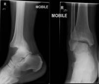
Figure 2.Lateral (left image) and AP (right image) radiograph of a posterior ankle dislocation (image courtesy of Dr. Henry Knipe from the Radiopaedia case on Ankle Fracture-Dislocation)
For uncomplicated ankle dislocations, there should be no additional fractures and the tibiofibular syndesmosis should not be pathologically widened. With regards to posterior dislocations, the talus is located posteriorly to the tibia and may be better visualized on lateral views (Figure 2). While plain films should be the initial imaging study, CT imaging should be considered to evaluate for concomitant injury or osteochondral fragments trapped within the joint space.5
The Bosworth fracture-dislocation is a rare entity, which is a posterior talar dislocation and displacement of the distal fibula posterior to the distal tibia, best noticed on the lateral xray. On plain film, it may initially appear as a posterior dislocation with a uni/bimalleolar fracture.7 However, it is extremely difficult to reduce as the proximal part of a fibular fracture becomes entrapped behind the lateral tibial tubercle. Reduction of the tibio-talar joint may be achieved in some circumstances; however, the fibular fragment will remain entrapped posteriorly. Given patient discomfort, positioning of lateral radiographs may not reveal the displacement. With a high degree of suspicion, CT imaging is recommended.7 Open reduction and internal fixation is necessary to achieve appropriate alignment.
Reduction Technique
Ensure the patient has received sedation and/or pain medications for comfort and to increase reduction success. Usually an assistant is needed to help with patient positioning and providing traction during the reduction procedure. When reducing any joint, the initial step is to recreate the mechanism. Knee flexion can help to remove tension on the Achilles tendon at the heel.5
Lateral fracture-dislocations2
Bend the knee 90°
Plantar-flex the ankle
Apply axial traction on the foot
Apply lateral traction proximal to and medial pressure distal to the injury
Posterior ankle dislocation2
Bend the knee 90°
Plantar-flex the ankle
Apply axial traction on the foot
Apply posterior traction proximal to and anterior pressure distal to the injury
Similar approaches are taken for medial and anterior ankle dislocations.
Splint
Patients should have a short leg posterior splint placed after reduction. A stirrup can provide additional stability.
Return Precautions & Post-Discharge Care
Similar to other lower extremity injuries, patients should be advised to keep their extremity elevated to reduce swelling. They should be given crutches and advised to be non-weight-bearing until re-evaluated by an orthopedist. They should be instructed regarding proper splint care and to monitor for new numbness, tingling, weakness, discoloration, or worsening pain for potential compartment syndrome. Decisions regarding DVT prophylaxis and anticoagulation should be made in conjunction with the orthopedist involved with the patient’s care.
When to Consult an Orthopedist in the Emergency Department
Depending on the extent of injuries and institutional culture, initial orthopedic consultation and follow up scheduling can vary. Patients with signs of neurovascular compromise or skin tenting from an ankle dislocation should be reduced immediately without waiting for radiographs or an orthopedics consultation. Below is a general list of when to involve orthopedics:
Associate Professor of EM, University of ArizonaAssociate Program Director, Sports Medicine Fellowship Team Physician for the University of Arizona
Neurovascular exam: Ankle dislocations typically occur due to a high mechanism force. They frequently have associated fractures and neurovascular (NV) injuries – it is imperative to perform a quick but thorough NV exam upon initial presentation and monitor that status throughout the ED course. Do not delay reduction for radiographs if there is compromise.
Soft tissue injury: Isolated dislocations can lead to significant instability due to ligamentous or cartilage injuries – there is a risk for chronic ankle instability.
Unable to reduce: If unable to reduce the dislocation in the ED, consider a Bosworth fracture, underlying cartilage disruption, or osteochondral injury. CT imaging is recommended.
Mimicker: Differentiate a true ankle (talar) dislocation from a subtalar dislocation, as the methods for reduction are different. Subtalar dislocations can be more difficult to reduce in the ED.
Post-reduction care: All patients with ankle dislocations being discharged require complete instructions including splint care, monitoring for complications, and close outpatient orthopedic follow-up. Ligaments are torn in dislocations and can sometimes result in chronic ankle stability.
References
Carragee EJ, Csongradi JJ, Bleck EE. Early complications in the operative treatment of ankle fractures. Influence of delay before operation. J Bone Joint Surg Br 1991;73(1):79–82. PMID 1991782
Handel DA, Gaines SA. Ankle Injuries. In: Tintinalli JE, Stapczynski JS, Ma OJ, Yealy DM, Meckler GD, Cline DM, editors. Tintinalli’s Emergency Medicine: A Comprehensive Study Guide, 8e. New York, NY: McGraw-Hill Education; 2016.
Davenport M. Ankle [Internet]. In: Sherman SC, editor. Simon’s Emergency Orthopedics. New York, NY: McGraw-Hill Education; 2014.
Schwartz DT. Chapter IV-1. Ankle Fractures. In: Emergency Radiology: Case Studies. New York, NY: The McGraw-Hill Companies; 2008.
Mubark I, Anwar S, Hayward K. Closed posterior ankle dislocation without associated fractures: a case report. J Surg Case Rep 2017 [cited 2019 Apr 13];2017(8). PMID 28928920
Agrawal AC, Raza H, Haq R. Closed posterior dislocation of the ankle without fracture. Indian J Orthop 2008;42(3):360–2. PMID 19753168
Sore throat accounts for a whopping 7.3 million outpatient pediatric visits. Group A Streptococcus (GAS) accounts for 20-30% of pharyngitis cases with the rest being primarily viral in etiology. However, clinically differentiating viral versus bacterial causes of pharyngitis is difficult and we, as providers, often don’t get it right. In addition, antimicrobial resistance is increasing.. So who do we test and when do we treat for strep throat? The 2012 Infectious Diseases Society of America (IDSA) guideline on GAS pharyngitis helps answer these questions.
Bacterial or viral pharyngitis?
Group A streptococcus (GAS) is the most common bacterial cause of pharyngitis in both children and adults. It is important to diagnosis and treat GAS pharyngitis to prevent the non-suppurative complication of acute rheumatic fever and suppurative complications such as peritonsillar abscess, retropharyngeal abscess, mastoiditis, and lymphadenitis.1 Additionally, it is important to rule out GAS pharyngitis so as to avoid unnecessary antibiotic use in a time of increasing antibiotic resistance patterns.
Strep pharyngitis most commonly occurs in children ages 5-15 years old, and symptoms include sore throat, pain with swallowing, and fever. In children, headache, nausea, abdominal pain, and vomiting are also commonly present. Physical exam findings include tonsillopharyngeal erythema and exudates, cervical lymphadenopathy, uvula swelling, palatal petechiae, and a scarlatiniform rash. Viral pharyngitis may present similarly to GAS pharyngitis; however, a lack of fever and the presence of rhinorrhea, cough, conjunctivitis, stomatitis, oral ulcers, and viral exanthem suggest a more viral etiology.
While there exist several prediction tools designed to aid in the clinical diagnosis of GAS pharyngitis, such as the Centor and McIsaac Criteria, none perform well in children.2,3 In some cases these scoring systems can help to identify children at low risk for GAS and therefore reduce the need for further testing; however, as many as 65% of patients who present with all of the clinical criteria in a particular tool will test negative for GAS on throat culture, indicating a viral etiology.1
Table 1: Classic symptoms and findings for a viral and bacterial pharyngitis
Symptom
Viral
Bacterial
Sore throat
+
+
Scarlatiniform rash
–
+
Fever
+/-
+
Pain with swallowing
+
+
Headache
+/-
+
Nausea/vomiting/abdominal pain
–
+
Rhinorrhea
+
–
Cough
+
–
Conjunctivitis
+
–
Stomatitis/oral ulcers
+
–
Viral exanthem
+
–
How to test: Rapid Antigen Detection Tests and
the strep culture
In patients with suspected GAS pharyngitis a Rapid Antigen Detection Test (RADT) should be used for diagnosis. RADTs allow providers to quickly test for GAS instead of relying on inadequate clinical tools or waiting for a strep culture to result. They are highly specific with a low false positive rate. Thus if positive, the patient should be treated with antibiotics and a confirmatory strep culture is not necessary. Of note, rapid strep testing will remain positive on average for 4 days after initial diagnosis, but can remain positive for up to 2 weeks depending on the individual and antibiotic compliance. Repeat testing with RADT after a course of antibiotics for GAS pharyngitis should be reserved for patients only with the recurrence of classic symptoms of strep throat.4
What if the
rapid strep test is negative?
RADTs have a sensitivity of 70-90%, leading to some false negative results.5,6 Thus if the RADT result is negative, a strep culture should be sent with a follow-up plan, should the culture become positive. Antibiotics can be initially withheld, unless the patient is at high risk (immunosuppressed, medically complex) or has high-risk contacts.
Who to test: Children younger than 3 years old
don’t need to be tested
GAS pharyngitis is rare (0-14%) in children <3 years of age.7 Furthermore, the incidence of rheumatic fever is rare.8 The 2012 IDSA guidelines recommended that routine testing for GAS pharyngitis in patients <3 years of age is NOT indicated. Only in situations of a household contact with known GAS infection would it be reasonable to consider testing.1
How to treat GAS pharyngitis
Fortunately GAS is a relatively easy bug to kill. It is susceptible to penicillins and its sister beta-lactams, amoxicillin, and ampicillin. While penicillin is cheaper and as efficacious as amoxicillin, pediatrician tend to choose a 10-day course of amoxicillin due to its better taste and therefore higher compliance rate.
For penicillin-allergic patients, first generation cephalosporins such as cephalexin are recommended in those without anaphylaxis to penicillins. For those with anaphylaxis to penicillins, a 10-day course of clindamycin or a 5-day course of azithromycin is recommended.1
Table 2: Antibiotic recommendations for Group A Streptococcal pharyngitis per 2012 IDSA guidelines, if the patient is NOT allergic to penicillin
Antibiotic
Dosing
Duration
Penicillin V
*Children: 250 mg po BID/TID *Adolescent/Adults: 250 mg po QID or 500 mg po BID
10 days
Amoxicillin
* 50 mg/kg (max 1,000 mg) po daily, or * 25 mg/kg (max 500 mg) po BID
10 days
Benzathine penicillin G
* Weight <27 kg: 600,000 units IM * Weight ≥27 kg: 1.2 million units IM
1 time dose
Table 3: Antibiotic recommendations for Group A Streptococcal pharyngitis per 2012 IDSA guidelines, if the patient IS allergic to penicillin (*avoid if anaphylactic to penicillin)
Antibiotic
Dosing
Duration
Cephalexin*
20 mg/kg/dose (max 500 mg) po BID
10 days
Cefadroxil*
30 mg/kg (max 1,000 mg) po daily
10 days
Clindamycin
7 mg/kg/dose (max 300 mg) po TID
10 days
Azithromycin
12 mg/kg (max 500 mg) po daily
5 days
Clarithromycin
7.5 mg/kg/dose (max 250 mg) po BID
10 days
What about GAS carriers?
GAS carriers are patients with persistent GAS positive throat cultures despite treatment and without any further symptoms of GAS pharyngitis. These patients have GAS present in the pharynx but no signs of immunologic response, meaning that their antistreptolysin O (ASO) titers are negative.9
RADTs and strep cultures do not distinguish between active infection and carriers. Carriers do not require treatment and have a low risk of spreading infection to those in close contact. They are also at low risk for developing suppurative and non-suppurative complications.
Can we do better?
Despite the fact that RADTs have the potential to drastically reduce the number of antibiotic prescriptions for viral pharyngitis, prescribing rates remain high. Studies cite that antibiotics are prescribed in as many as 53% of all patients with pharyngitis symptoms, which is, well above the known prevalence of GAS at 20-30%.8 So why are we still giving antibiotics for viral pharyngitis? The answer is probably multifactorial, including providers empirically treating sore throat without testing, testing in inappropriate cases, such as young children, and the increasing prevalence of carrier states.
Take Away Points
Do not rely on the clinical diagnosis for GAS pharyngitis in children. Instead use a Rapid Antigen Detection Test (RADT) and, if negative, a throat culture for diagnosis.
There is no indication to test children <3 years of age for GAS pharyngitis with the RADT or strep culture unless there is a known household contact with GAS.
Treat with a 10 day course of amoxicillin or cephalexin in non-anaphylactic, penicillin-allergic patients. Clindamycin or azithromycin are appropriate antibiotics in anaphylactic, penicillin-allergic patients.
References
1.
Shulman S, Bisno A, Clegg H, et al. Clinical practice guideline for the diagnosis and management of group A streptococcal pharyngitis: 2012 update by the Infectious Diseases Society of America. Clin Infect Dis. 2012;55(10):1279-1282. https://www.ncbi.nlm.nih.gov/pubmed/23091044.
2.
Shaikh N, Swaminathan N, Hooper E. Accuracy and precision of the signs and symptoms of streptococcal pharyngitis in children: a systematic review. J Pediatr. 2012;160(3):487-493.e3. https://www.ncbi.nlm.nih.gov/pubmed/22048053.
3.
Fine A, Nizet V, Mandl K. Large-scale validation of the Centor and McIsaac scores to predict group A streptococcal pharyngitis. Arch Intern Med. 2012;172(11):847-852. https://www.ncbi.nlm.nih.gov/pubmed/22566485.
4.
Homme J, Greenwood C, Cronk L, et al. Duration of Group A Streptococcus PCR positivity following antibiotic treatment of pharyngitis. Diagn Microbiol Infect Dis. 2018;90(2):105-108. https://www.ncbi.nlm.nih.gov/pubmed/29150372.
5.
Tanz R, Gerber M, Kabat W, Rippe J, Seshadri R, Shulman S. Performance of a rapid antigen-detection test and throat culture in community pediatric offices: implications for management of pharyngitis. Pediatrics. 2009;123(2):437-444. https://www.ncbi.nlm.nih.gov/pubmed/19171607.
6.
Gerber M, Shulman S. Rapid diagnosis of pharyngitis caused by group A streptococci. Clin Microbiol Rev. 2004;17(3):571-580, table of contents. https://www.ncbi.nlm.nih.gov/pubmed/15258094.
7.
Nussinovitch M, Finkelstein Y, Amir J, Varsano I. Group A beta-hemolytic streptococcal pharyngitis in preschool children aged 3 months to 5 years. Clin Pediatr (Phila). 1999;38(6):357-360. https://www.ncbi.nlm.nih.gov/pubmed/10378093.
8.
Tani L, Veasy L, Minich L, Shaddy R. Rheumatic fever in children younger than 5 years: is the presentation different? Pediatrics. 2003;112(5):1065-1068. https://www.ncbi.nlm.nih.gov/pubmed/14595047.
9.
Johnson D, Kurlan R, Leckman J, Kaplan E. The human immune response to streptococcal extracellular antigens: clinical, diagnostic, and potential pathogenetic implications. Clin Infect Dis. 2010;50(4):481-490. https://www.ncbi.nlm.nih.gov/pubmed/20067422.
Author information
Shannon Flood, MD
Pediatric Emergency Medicine Fellow
Children's Hospital Colorado
Severe constipation, requiring fecal disimpaction and rectal enemas, can be excruciatingly painful for patients. Administering sedatives and opioids to help alleviate this pain poses a challenge, because many of the patients are elderly and tend to be more sensitive to these medications. Furthermore, there may be increased vagal tone when straining, leading to hypotension and bradycardia and which can result in defecation-related syncope. 1 Also, opioids can exacerbate constipation. Herein we present 2 cases and tricks on achieving better pain control.
Case 1: Painful disimpaction
A 75-year-old man presents to the Emergency Department (ED) with severe constipation. He reports having no bowel movements for a week, despite multiple laxatives and 2 Fleets enemas. The provider suspects fecal impaction and the rectal exam confirms this suspicion. The provider prepares for digital disimpaction, which is often a painful procedure.
Trick of the Trade Use topical 2% lidocaine jelly (10 mL Sterile Pak Uro-Jet) to provide rectal lubrication as well as topical anesthesia. It is pre-dosed and in an applicator that makes rectal insertion simple. Administer all 10 mL of the jelly 5 minutes prior to the disimpaction procedure for the lidocaine to take effect. Hopefully with better pain control, manually break up the hard stool and remove it with a gloved index finger in a slightly bent, “hooking” fashion.2
Case 2: Painful rectal enemas
A 59-year-old woman presents to the ED with severe constipation. She reports that her stools have been scant and hard, but now she is having significant rectal pain when trying to have a bowel movement. She self-administered a Fleets enema prior to arrival, but it was too painful to expel the stool. You attempt a soap suds or milk and molasses enema in the ED, but the patient is retaining the stool and enema secondary to peri-rectal pain.
Trick of the Trade Administer an enema slowly as tolerated by the patient. Subsequently administer 10 mL of 2% lidocaine jelly rectally after the patient has retained the enema for about 20 minutes. The lidocaine provides lubrication and topical anesthesia to the rectal area, facilitating easier passage of the stool and enema.
Caution: Lidocaine systemic absorption
Lidocaine absorption via mucous membranes is varied and the extent depends on the concentration and the amount administered. Avoid repeated doses in short intervals. Avoid use in sepsis or severely traumatized mucosa since this could result in more systemic absorption. The maximum recommended dose of lidocaine is 4.5 mg/kg.
Wenzke K, Walsh K, Kalscheur M, et al. Clinical Characteristics and Outcome of Patients with Situational Syncope Compared to Patients with Vasovagal Syncope. Pacing Clin Electrophysiol. 2017;40(5):591-595. https://www.ncbi.nlm.nih.gov/pubmed/28244210.
Instructor of Emergency Medicine
Mayo Clinic, Jacksonville, Florida;
Clinical Assistant Professor
Lead Faculty Emergency Nurse Practitioner program
Jacksonville University
History of Present Illness: Patient is a 35-year-old transgender male with a history of bipolar disorder (taking seroquel/lamotrigine) who presents with 2 days of:
Flu-like symptoms
Progressive lip pain/swelling
Mouth pain
Oral ulcers
Eye redness
New erythematous rash involving the palms/soles and lower extremities
The patient initially noted myalgias, fever, and malaise 2 days ago. Yesterday, the patient woke up with bilateral eye redness and itching, and he developed lip swelling/discoloration and mouth pain throughout the day. He presented to an outside emergency department (ED) 12 hours prior, where he was told that he had a viral infection, given pain medication, and discharged home. He has not taken any other medications. The patient presents to this ED due to progression of symptoms, including the development of a pruritic rash on his palms, soles, and lower extremities. Upon further questioning, the patient also reports vaginal itching and a fishy odor. He has a history of bacterial vaginosis and states that these symptoms feel similar. The patient denies genital sores, vaginal discharge, and vaginal bleeding. He is currently sexually active with men and women, and does not regularly use barrier protection.
Mouth: Dusky/purple lips with a bright erythematous rim around the vermillion border, multiple ~5mm ulcerations of the anterior oral mucosa, erythema of hard and soft palates with several punctate erosions
Neck: Tender cervical lymphadenopathy
Skin: Multiple 2-3mm erythematous papules on the bilateral palms, soles and thighs; no vesicles or bullae, no skin sloughing
This patient was ultimately diagnosed with EM Major, confirmed with skin biopsy.
EM is a type IV hypersensitivity reaction to infections (commonly HSV or mycoplasma) or medications (including sulfonamides and hydantoins).
The classic targetoid papules are often preceded by malaise, fever, and itching at the site where eruptions later occur. EM exhibits less mucous membrane and body surface area involvement compared to Stevens-Johnson syndrome and toxic epidermal necrolysis.
Herpes simplex virus (HSV) is the most common etiology of erythema multiforme, accounting for more than 50% of cases. Early initiation of oral acyclovir may lessen the severity of cutaneous lesions.
Mild cases of erythema multiforme can be managed with oral antihistamines and topical steroids. More severe cases may require hospitalization and oral or intravenous steroid administration. In all cases, the underlying cause should be addressed if possible by treating the infectious cause or discontinuing the causal medication.
Copyright
Images and cases from the Society of Academic Emergency Medicine (SAEM) Clinical Images Exhibit at the 2019 SAEM Annual Meeting | Copyrighted by SAEM 2019 – all rights reserved.
Author information
Nicholas Stark, MD
Emergency Medicine Resident
University of California San Francisco
History of Present Illness: An 89-year-old female with a past medical history of coronary artery disease and with recent admission for myocardial infarction that was medically managed, presented with chest pain and shortness of breath. She reports worsening midsternal chest pain that occasionally radiates to her back and right arm since discharge.
Figure 2: Patient ECG
Figure 3: Patient bedside cardiac ultrasound with color Doppler
Figure 1 shows a circumferential pericardial effusion.
The ECG (figure 2) shows a lateral myocardial infarction, which is likely the progression of her myocardial infarction that was medically managed previously.
Figure 3 image shows the free wall rupture with a “jet” of blood leaving the lateral wall using Color Doppler.
Left ventricular free wall rupture happens in about 2% of all patients with acute myocardial infarction.
Rupture typically occurs about 3-5 days after infarction.
There are 3 classifications, Type I–III, which are dependent on the time of rupture from when the infarction occurred. Rupture occurs near the edge of where necrotic myocardium meets healthy myocardium. Treatment for left ventricular rupture is supportive with blood pressure management in the immediate setting. If the patient does survive acute rupture, surgical correction can be performed if feasible. Prognosis is dependent on multiple factors, but overall prognosis is poor with mortality near 100%.
This patient was diagnosed with left ventricular free wall rupture after bedside ultrasound was performed. The patient’s previous myocardial infarction happened four days prior to presentation. The patient survived her weeklong hospital stay and elected for medical management only. The patient was discharged home in stable condition.
Copyright
Images and cases from the Society of Academic Emergency Medicine (SAEM) Clinical Images Exhibit at the 2019 SAEM Annual Meeting | Copyrighted by SAEM 2019 – all rights reserved.
Author information
Andrew Ortega, MD
Clinical Assistant Professor
Department of Emergency Medicine
Kaiser Permanente Central Valley
One of the gold standard for building and sustaining collaborative, multi-institutional research networks in medicine is the Pediatric Emergency Care Applied Research Network (PECARN) organization. Their efforts on studying pediatric emergency care has resulted some of our specialty’s landmark papers in Lancet, New England Journal of Medicine, JAMA Pediatrics, and Annals of Emergency Medicine. Although we are not officially affiliated with them, we fully support their efforts and wanted to help disseminate their evidence-based findings with an educations. Thus was born the PECARN Publication Prospectus (P3) app project [download free P3 app].
The P3 Project and Team
As with many of our ALiEM initiatives, the P3 project arose from a collaborative sprint effort over a 4 week period in 2019 with prehospital educators, emergency medicine (EM) residents, budding and current pediatric EM fellows, and EM/PEM attending physicians. This app plans to be a “living” catalog of PECARN publications which is updated as their prolific research team continues to publish.
Phase 1: Extracting the clinically-relevant educational pearls and a brief study summary from each of their 140+ peer-reviewed papers
Phase 2: Feature expert peer-reviewer commentaries from one of the paper’s authors
Phase 3: Link high-quality online resources which review or highlight these papers
Paramedic/ Firefighter, Montgomery Co. Fire-EMS; Critical Care Flight Paramedic, U.S. Army National Guard
Ginger Locke, BA NRP
Associate Professor of EMS Professions, Austin Community College
Floyd Miracle, BS NRP
Clinical Manager, Jessamine County EMS
Damian Roland, BMedSci, BMBS, PhD
Honorary Associate Professor and Consultant in Paediatric EM
Jason Woods, MD
Assistant Professor of Pediatrics, University of Colorado, Children’s Hospital of Colorado
Michelle Lin, MD
ALiEM Founder; Professor of EM, UC San Francisco
P3 App
The P3 app, which is compatible with iOS and Android devices, summarizes each of the 140+ PECARN publications. These papers are subcategorized into learner groups (physicians/advanced practice providers, pharmacists, triage nurses, prehospital providers, and administrators) as well as organ system groups.
We are always looking for more volunteers (physicians, pharmacists, nurses, paramedics) who support this exciting initiative. Contact us at info@aliem.com. Specifically, we need assistance with:
Designing a P3 logo
Identifying high-quality online resources that discuss the PECARN publications as part of Phase 3
History of Present Illness: The patient is an 18 year-old male who presents with a rash that appeared 7 days ago. The rash is located on his torso, back, and lower lip. It is pruritic. Three days prior to the appearance of the rash, he had a sore throat and intermittently took ibuprofen over the ensuing 3 days. He stopped taking ibuprofen 4 days after his sore throat abated. He denies any fever, nausea, vomiting, shortness of breath, chest pain, abdominal pain, diarrhea, extended travel in the past year, sick contacts, new soaps/detergents, insect bites, chemical exposure, and new foods.
HEENT: Unremarkable oropharynx without mucosal involvement
Pulmonary: Lungs clear to auscultation bilaterally
Abdominal: Abdomen is soft and non-tender
Skin: There are several dozen raised, hyperpigmented circular lesions 1-1.5 inches in diameter on the patient’s torso and back. The borders are sharply demarcated. Some of the lesions appear to be fluid-filled. Nikolsky sign is negative.
Pathology report: “Parakeratosis, dyskeratotic keratinocytes, and vacuolar interface dermatitis in the superficial epidermis with prominent pigment incontinence.”
The photos depict a Fixed Drug Eruption (FDE). FDE is characterized by oval, raised, well-demarcated, and hyperpigmented lesions. The diagnosis is supported by a recent history (hours to days) of exposure to NSAIDs.
Always consider a Fixed Drug Eruption when a patient reports recent NSAID exposure and presents with a diffuse pruritic rash consisting of circular lesions.
Copyright
Images and cases from the Society of Academic Emergency Medicine (SAEM) Clinical Images Exhibit at the 2019 SAEM Annual Meeting | Copyrighted by SAEM 2019 – all rights reserved. View other other cases from this series on ALiEM.
Author information
Zachary Smith, MD
PGY-II House Officer
Department of Emergency Medicine
Wake Forest Baptist Medical Center
The skilled and rapid resuscitation of critically ill patients is a central premise in the specialty of emergency medicine (EM). A paradox for providers often arises when in the midst of resuscitating a patient with advanced chronic illness, the question of risks versus benefits arises. For this patient, we may successfully stabilize vital signs, but at what cost? Will this patient return to a quality of life they deem acceptable? What are the patient’s goals of treatments given his/her underlying disease? These questions illustrate the need for emergency physicians to be more aware of and comfortable with palliative care practices.
“… an approach that improves the quality of life of patients and their families who are facing problems associated with life-threatening illness. It prevents and relieves suffering through the early identification, correct assessment and treatment of pain and other problems, whether physical, psychosocial, or spiritual.”
As a “front-door” medical specialty for a variety of conditions and injuries across age groups, the emergency department (ED) can also serve as the front door to palliative care. We witness first-hand the acute presentations of infection, respiratory distress, neurologic compromise, and hemodynamic instability secondary to these illnesses. These acute changes in the disease course can serve as an opportunity to revisit goals and next steps in an effort to ensure that the care initiated by emergency providers is concordant with the patient’s own goals. These conversations have the potential to change the trajectory of the patient’s course of care.1–3
However, we emergency physicians are often not trained for this.4 There has been a lack of education on palliative care topics in the EM residency curriculum as the focus of EM has traditionally been to prolong life. As a result, we have developed a culture in which “success” is equated to “avoiding death.” We are rarely asked to consider when we are prolonging the dying process, and not the living.5
Palliative emergency medicine, defined as the integration of palliative care principles into emergency medicine practice, places the patient back at the center of care, eliciting the person’s own values, concerns, and decisions. It enables the physician to support those goals through clear communication, easing suffering, and creating a partnership. While not meant to replace the central premises of EM, incorporating palliative care skills into one’s armamentarium is meant to supplement and assist providers in providing care that is truly aligned with the patient and family. It is this shared partnership that may also be the key to physician resilience and longevity.6
Benefits for our emergency department patients
Palliative care is a truly patient-centered approach to care. There is no one definition of good quality of life. This is a dynamic process that is negotiated and re-negotiated amongst patients, families, and health care professionals, framed by individual values, knowledge, and preferences for care.7
What are the benefits of palliative care?1
Improved satisfaction for patients and families
Improved symptom management
Less time in the intensive care unit
Increased appropriate use of hospice
Better resource management
Initiating a palliative care consultation directly from the ED (versus later as an inpatient) shortens length of stay by an average of 4 days, resulting in fewer in-hospital deaths while significantly increasing quality of life, without reducing overall survival.8
A 2010 landmark study also reveals what many may consider surprising. Early palliative care intervention in patients with metastatic cancer increased length of survival by 3 months in a group with early palliative intervention and standard oncologic care, as compared to standard oncologic treatment alone, despite the fact that fewer patients in the early palliative care group received aggressive end-of-life care.9
Benefits for us as EM providers: Protective effects against burnout
Burnout rates for emergency providers can be as high as 65%, which is more than any other specialty and is seemingly increasing [ACEP News, 2017]. Our setting is ripe for emotional exhaustion and depersonalization given the nature of high acuity and overwhelming volume of patients needing care.
Palliative care encompasses many of ideals, which have been noted to protect against compassion fatigue and burnout.
Emotional resilience: Resiliency of the human spirit and the opposite of emotional exhaustion10
The burnout phenomenon may be viewed as a result of a movement away from bedside care toward a more facile business-like knowledge of systems which enable “efficiency” within these systems. We may have moved from an experience of connections with patients to one of external facility. In this process, we have moved away from the deep satisfaction of what is non-physical about caring for patients. Building awareness of the emotional and spiritual needs of patients has a positive effect on both those providing the care and those receiving it.11
Palliative care offers the possibility to re-engage with patients about what is most important to them, placing the patient again at the center of care. What many of us seek in becoming physicians is a reciprocal healing process, which occurs through meaningful relationships, inner self-reflection, and a connection with peers and community.10 Palliative care offers this engagement, and hence offers us an ability to find our own emotional resilience.
With a bird’s-eye view of the ED, it may not seem the ideal place for intimate questions about life and death. However, the ED bedside may be the richest place to do so, engaging the patient and family soon after s/he enters the hospital, in a vulnerable moment during an acute presentation, before his/her trajectory is set in motion. Here we may also find the keys to our own personal re-engagement as providers, with physicians and patients working together in a shared process, communicating about values and short- and long-term goals.
Call to action: Incorporate palliative care into EM practices
As emergency physicians, we can be the catalyst in a patient’s journey to begin crucial palliative care conversations with patients and their families. Consider tackling questions such as, “What is the meaning of living well for you?” In these conversations, we may uncover our own well-spring of emotional resilience. For many reasons, this conversation is perhaps one of our most relevant critical procedures in the ED.8
Grudzen C, Stone S, Morrison R. The palliative care model for emergency department patients with advanced illness. J Palliat Med. 2011;14(8):945-950. https://www.ncbi.nlm.nih.gov/pubmed/21767164.
4.
Quest T, Asplin B, Cairns C, Hwang U, Pines J. Research priorities for palliative and end-of-life care in the emergency setting. Acad Emerg Med. 2011;18(6):e70-6. https://www.ncbi.nlm.nih.gov/pubmed/21676052.
Steinhauser K, Christakis N, Clipp E, McNeilly M, McIntyre L, Tulsky J. Factors considered important at the end of life by patients, family, physicians, and other care providers. JAMA. 2000;284(19):2476-2482. https://www.ncbi.nlm.nih.gov/pubmed/11074777.
Temel J, Greer J, El-Jawahri A, et al. Effects of Early Integrated Palliative Care in Patients With Lung and GI Cancer: A Randomized Clinical Trial. J Clin Oncol. 2017;35(8):834-841. https://www.ncbi.nlm.nih.gov/pubmed/28029308.
10.
Kearney M, Weininger R, Vachon M, Harrison R, Mount B. Self-care of physicians caring for patients at the end of life: “Being connected… a key to my survival”. JAMA. 2009;301(11):1155-1164, E1. https://www.ncbi.nlm.nih.gov/pubmed/19293416.
11.
Harrison R, Westwood M. Preventing vicarious traumatization of mental health therapists: Identifying protective practices. Psychotherapy (Chic). 2009;46(2):203-219. https://www.ncbi.nlm.nih.gov/pubmed/22122619.
Author information
Beth Krafchik, MD
PGY-4 Resident Physician
Department of Emergency Medicine
Kings County Hospital Center/SUNY Downstate Medical Center
Welcome to the AIR Orthopedics Upper Extremity Module! After carefully reviewing all relevant posts from the top 50 sites of the Social Media Index, the ALiEM AIR Team is proud to present the highest quality online content related to orthopedics upper extremity emergencies. 5 blog posts within the past 12 months (as of March 2019) met our standard of online excellence and were curated and approved for residency training by the AIR Series Board. We identified 0 AIR and 5 Honorable Mentions. We recommend programs give 2.5 hours (about 30 minutes per article) of III credit for this module.
AIR Series Stamp of Approval and Honorable Mentions
In an effort to truly emphasize the highest quality posts, we have 2 subsets of recommended resources. The AIR stamp of approval is awarded only to posts scoring above a strict scoring cut-off of ≥30 points (out of 35 total), based on our scoring instrument. The other subset is for “Honorable Mention” posts. These posts have been flagged by and agreed upon by AIR Board members as worthwhile, accurate, unbiased, and appropriately referenced despite an average score.
Interested in taking the quiz for fun or asynchronous (Individualized Interactive Instruction) credit? Please go to the above link. You will need to create a free, 1-time login account.
Highlighted Quality Posts on Orthopedics Upper Extremity Emergencies
From the ALiEM AIR Executive Board and ALiEMU Team
Farhad Aziz
Jeremy Branzetti
Hari Bhatt
Chris Belcher
Adam Evans
Sean Fox
Chris Gaafary
Andrew Grock
Jacob Hennings
Jaime Jordan
Nikita Joshi
Jay Khadpe
Michelle Lin
Allie Min
Eric Morley
Salim Rezaie
Lynn Roppolo
Matthew Rosen
Kaushal Shah
Derek Sifford
Anand Swaminathan
Wes Trueblood
Natasha Wheaton
David Yang
Author information
Chris Belcher, MD
Co-Editor, ALiEM AIR Series
Assistant Professor and Staff Physician
Department of Emergency Medicine
San Antonio Uniformed Services Health Education Consortium
Supracondylar humerus fractures are the most common type of elbow fracture in pediatric patients, most often seen in a fall on an outstretched hand (FOOSH) or a fall on a hyper-extended elbow.1,2 If there is no obvious fracture on x-rays, the patient may have an occult fracture; look for secondary radiographic signs including a posterior fat pad sign, an enlarged anterior fat pad or ‘sail sign’, or malalignment. Occult supracondylar fractures (those with initial normal radiographs that are later diagnosed in follow up) make up 2-18% of all the fractures we see in kids.3 When x-ray findings are nonspecific but the index of suspicion for fracture remains high, ultrasound may aid in your clinical decision making.
Technique
Probe: High resolution linear probe with frequencies ranging from 6-13 MHz
Maximum Depth:6 cm
Scanning Scheme: With the elbow bent at 90 degrees, scan along posterior edge of the distal humerus in longitudinal and transverse axes.
Findings on Elbow Ultrasound
Normal Elbow
Identify:
Distal humerus without cortical irregularity
Posterior fat pad
Abnormal Elbow
Identify signs of potential occult fracture:
Bulging posterior fat pad
lipohemarthrosis
Elbow Ultrasound vs Radiograph
Study 1: European Journal of Trauma and Emergency Surgery 2014
A German study investigated ultrasound accuracy (identifying cortical deformity or a posterior fat pad) compared to standard radiographs in diagnosing supracondylar fractures in 106 pediatric patients. The test characteristics were as follows:4
Sensitivity 100%
Specificity 93.5%
Negative predictive value 100%
Positive predictive value 95.2%
Study 2: Orthopaedics & Traumatology: Surgery & Research 2016
A French study of 34 cases of pediatric elbow trauma with suspected occult fracture. Patients with bone deformity diagnosed on initial radiographs were excluded. Patients were splinted until they had an ultrasound performed within 6 days of the injury.3
13 of the 34 cases had identified an occult fracture
Of the 21 cases who had normal ultrasounds, none were later diagnosed with a fracture.
Posterior fat pad sign sensitivity 100% (13 cases)
Lipohemarthrosis sensitivity 92% (11 cases)
Cortical disruption (11 cases)
Study 3: Annals of Emergency Medicine 2013
130 pediatric patients with elbow injuries. Before obtaining radiographs, pediatric emergency physicians performed an ultrasound. 33% patients had a radiograph result positive for fracture.5The ultrasound demonstrated either an elevated posterior fat pad or lipohemarthrosis with the following test characteristics:
Sensitivity 98%
Specificity 70%
Positive likelihood ratio 3.3
Negative likelihood ratio 0.03
Study 4: Pediatric Radiology 2008
14 patients with x-rays showing elbow joint effusion but no fracture who underwent ultrasound and MRI.6 The test characteristics for ultrasound in demonstrating lipohemarthrosis are as follows:
Sensitivity 88%
Specificity 100%
Negative predictive value 86%
Positive predictive value 100%
The Bottom Line
Elbow ultrasound can assist in ruling out a supracondylar fracture, which is particularly helpful in the case of an equivocal x-ray but high clinical suspicion.
Next time you encounter a pediatric elbow injury with negative x-rays in the ED, grab an ultrasound probe to evaluate for a fat pad sign and lipohemarthrosis.
Although not ready for prime time yet, this is an interesting technique that may result in decreased painful immobilization and improved overall morbidity. For now, continue to splint patients for whom you have high suspicion of an occult fracture.
Burnier M, Buisson G, Ricard A, Cunin V, Pracros J, Chotel F. Diagnostic value of ultrasonography in elbow trauma in children: Prospective study of 34 cases. Orthop Traumatol Surg Res. 2016;102(7):839-843. https://www.ncbi.nlm.nih.gov/pubmed/27697406.
4.
Eckert K, Janssen N, Ackermann O, Schweiger B, Radeloff E, Liedgens P. Ultrasound diagnosis of supracondylar fractures in children. Eur J Trauma Emerg Surg. 2014;40(2):159-168. https://www.ncbi.nlm.nih.gov/pubmed/26815896.
5.
Rabiner J, Khine H, Avner J, Friedman L, Tsung J. Accuracy of point-of-care ultrasonography for diagnosis of elbow fractures in children. Ann Emerg Med. 2013;61(1):9-17. https://www.ncbi.nlm.nih.gov/pubmed/23142008.
6.
Zuazo I, Bonnefoy O, Tauzin C, et al. Acute elbow trauma in children: role of ultrasonography. Pediatr Radiol. 2008;38(9):982-988. https://www.ncbi.nlm.nih.gov/pubmed/18626636.
Author information
Winnie W. Chan, MD
Chief Resident
UCSF-ZSFGH Emergency Medicine Residency Program
Department of Emergency Medicine
University of California, San Francisco
A 25-year-old medical student comes in with a muffled voice, sore throat and trismus. You look at the back of her throat and you see the uvula deviated to the right. You astutely diagnosed a peritonsillar abscess (PTA). You consider aspirating and want to check for tips on how to successfully do this.
Using the intracavitary probe provides great views of both the abscess and the nearby vessels (figure 1). Not only does it confirm the presence of abscess vs. a phlegmon, POCUS also gives the operator an idea of the depth of the carotid artery with respect to the visible surface of the posterior pharynx and the TPA.
Figure 1. Ultrasound of a peritonsillar abscess with the carotid artery (red) posterior to the mass (measured by green lines) using the intracavitary probe. Photo courtesy of Dr. Viveta Lobo.
2. Visualization
Use the bottom part of a disposable light-affixed pelvic speculum with the top removed (figure 2), leaving just one piece that the patient can hold down.
Figure 2. OPB Medical ER-SPEC lighted speculum (or equivalent) with the top removed for the PTA drainage procedure
Pro tip #1: Note that by asking the patient to hold the light, the operator frees an extra hand for suction or stability of the other hand. Take the extra step setting up with sufficient lighting to optimize success.
Figure 3. The patient is given the speculum, direct Macintosh laryngosope blade, or video laryngoscope blade and instructed to depress the base of tongue (left). In the patient’s other hand holds a suction Yankauer (right).
3. Analgesia
Good analgesia is important for this anxiety-provoking and painful procedure. We have seen success with first using topical benzocaine spray. Alternatively, nebulizing lidocaine (2% or 4% without epinephrine) over 5 minutes can be used. Next, inject 2-3 mL of 1% lidocaine with epinephrine into the mucosa and muscle. 1
Pro tip #2: We expect the patient to salivate, or for pus to drain upon fenestration. Hand the patient the suction. Just like in the dental office, the patient can spit into the suction to optimize the view of the posterior pharynx, while minimizing risk for aspiration.
Pro tip #3: Communication is key. As it will be difficult for the patient to articulate concerns while you have the needle in hand and speculum retracting the tongue down, instruct the patient to lift the suction up to get your attention instead of saying something, or worse, pulling away. This ensures safety for the operator and the patient.
4. Drainage: Pediatric bullet tube as a depth guard
Many techniques exist for the actual drainage, including using a spinal needle or a traditional 1-1.5 inch needle, cutting the needle cover to buffer the depth of the actual needle. This protects the needle from penetrating the mucosa too deeply. Notice that the edge of the needle cover can be sharp and jagged. There is also a possibility for the plastic to chip off, thereby causing aspiration (Figure 4).
Figure 4. Cut the needle cover, exposing only 1 cm of the actual needle tip (left). The needle cover edge may be sharp and jagged (right).
Pro tip #4. We propose an alternative approach to a trimmed needle cover sheath. Cut the top lid off a pediatric bullet tube/microtainer and insert the 18g needle through the top onto the center part of the bottom aspect of the microtainer (figure 5). 1 cm of the needle will jut out of the back end of the tube. The cut bullet tube does not leave any sharp edges. The smooth surface of the back end of this microtainer, which will avoid traumatizing the posterior pharynx upon contact, or cause pieces of plastic to dislodge.
Figure 5.Cut the top lid off a pediatric bullet tube/microtainer (left) and use it to cover the 18g needle (right).
5. Aspiration
Aim the needle towards the area of most fluctuance.
Pro tip #5: Start with a 5-10 mL syringe instead of 20-30 mL syringe. This is because the larger the caliber of the syringe, the higher tension required to overcome the fluid pressure (remember LaPlace’s Law! Tension = pressure x radius), which may not be necessary in the setting of a fairly small abscess. Also, the smaller the syringe, the better access you have to the back of the patient’s throat.
Figure 6.Use 5-10 mL syringe to access easily the oropharynx. Notice the safety cover is pointed to the side to avoid this piece of plastic hitting the upper dentition upon entry.
Pro tip #6: While precision is key and aimed at the point of maximal fluctuance, multiple deliberate attempts actually leads to fenestrations that further allow for the pus to continue to drain post-procedure.
Using the operator’s dominant hand, grab the syringe with between the 1st and 2nd digits. As soon as the needle enters the mucosa, wrap the third digit around the plunger and apply negative pressure/pull back while advancing the syringe.
Figure 7. Single-handed aspiration of the syringe
6. Incision
Consider performing an incision and drainage (I&D) even after aspiration with the needle and syringe for large/loculated abscesses. A 2016 Cochrane review of I&D vs. needle aspiration found low quality evidence that I&D may have lower rates of recurrence than needle aspiration.2 We have found in practice that aspiration is great for removing pus but for large or loculated abscesses a single incision with 11-blade scalpel after the aspiration allows any remaining pus to drain and thus avoid recurrence.
Pro tip #7: Tape the cover of the scalpel/retractor to the desired depth. This ensures the scalpel will not penetrate more than 1 cm. In a single deliberate approach, incise about 1 cm along the area of maximal fluctuance (figure 8).
Figure 8. Scalpel with taped cover to avoid too deeply incising the abscess
Pro tip #8: Give the patient an ice cold water to swish and spit. It further helps control the pain, and allows for drainage to continue.
Pro tip #9: Put gloves on your patient’s dominant hand. The patient’s non-dominant hand is already holding the Yankauer suction (Figure 2). Instruct the patient to reach the newly drained abscess with their index finger, and ask the patient to place gentle pressure, “milking” the remainder fluid collection. The benefit of the patient doing this is:
It doesn’t trigger their gag reflex as much as it would if the operator does this.
The patient is less likely to bite their own hand.
The pressure placed on the back actually will help alleviate some of the pain they may be experiencing.
7. Procedural success
Successful drainage is seen when:
Fluctuance is less.
Patient’s trismus significantly improves.
Patient subjectively feels much better.
8. Post I&D care
PTA drainage leads to decreased oral intake, so most of these patients will be dehydrated. IV hydration may help with the patient subjectively feeling better. Oral antibiotics are always given, targeting Group A strep and oral anaerobes for 14 days.3 We suggest either regimen:
Amoxicillin-clavulanate 875 mg PO twice daily (45 mg/kg/dose for children up to maximum of 875 mg per dose), or
Clindamycin 300-450 mg PO every 6 hours (10 mg/kg/dose every 8 hours for children up to a maximum 450 mg per dose, although DynaMed/UpToDate states that 600 mg per dose is allowed), if MRSA is suspected.
Because of new emergence of antibiotic resistance, a culture from the abscess may be collected.4 Steroids also helps with alleviating trismus and can also reduce the pain and time of recovery.5 We suggest the regimen:
Dexamethasone 10 mg IV or PO
Case Conclusion
The patient feels significantly better. The muffled voice improved along with the trismus. The patient is given return precautions, emphasizing symptoms such as difficulty breathing, worsening pain or trismus, difficulty tolerating oral hydration, enlarging mass, fever, neck stiffness, or bleeding. The patient is discharged home with the antibiotic regimen above plus a recommendation for over-the-counter pain medications and instruction to gargle salt water.6
References
1.
Riviello R. Otolaryngologic Procedures. In: Roberts and Hedges’ Clinical Procedures in Emergency Medicine. Elsevier; 2014:1303-1309.
2.
Chang B, Thamboo A, Burton M, Diamond C, Nunez D. Needle aspiration versus incision and drainage for the treatment of peritonsillar abscess. Cochrane Database Syst Rev. 2016;12:CD006287. https://www.ncbi.nlm.nih.gov/pubmed/28009937.
3.
Kieff D, Bhattacharyya N, Siegel N, Salman S. Selection of antibiotics after incision and drainage of peritonsillar abscesses. Otolaryngol Head Neck Surg. 1999;120(1):57-61. https://www.ncbi.nlm.nih.gov/pubmed/9914550.
4.
Sowerby L, Hussain Z, Husein M. The epidemiology, antibiotic resistance and post-discharge course of peritonsillar abscesses in London, Ontario. J Otolaryngol Head Neck Surg. 2013;42:5. https://www.ncbi.nlm.nih.gov/pubmed/23663820.
5.
Chau J, Seikaly H, Harris J, Villa-Roel C, Brick C, Rowe B. Corticosteroids in peritonsillar abscess treatment: a blinded placebo-controlled clinical trial. Laryngoscope. 2014;124(1):97-103. https://www.ncbi.nlm.nih.gov/pubmed/23794382.